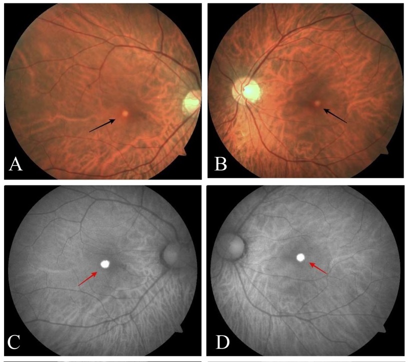
Pseudovitelliform maculopathy associated with hereditary hemochromatosis
Medical hypothesis discovery and innovation in ophthalmology,
Vol. 12 No. 4 (2023),
31 January 2024
,
Page 203-212
Abstract
Background: Hereditary hemochromatosis (HH) is an inherited autosomal recessive iron metabolism disorder resulting from a C282Y mutation in the HFE gene. Mutations in the HFE gene may result in iron accumulation and oxidative stress in the retina, resulting in macular degeneration. This article describes two patients with HH who were treated with erythrocytapheresis or phlebotomy, with no exposure to deferoxamine or any other chelation therapy, and who developed visual symptoms.Case Presentation: Both patients had known diagnoses of HH. Because of visual symptoms, they were referred to the ophthalmology clinic and underwent a retinal exam, multimodal imaging, and electrodiagnostic studies, which revealed structural and functional degeneration of the central macula. Fundus photography, fluorescein angiography, and fundus autofluorescence revealed changes at the level of the retinal pigment epithelium (RPE) in the central macula. In addition, optical coherence tomography revealed subfoveal accumulation of hyperreflective material at and below the RPE. Multifocal electroretinography confirmed a decreased cone response, whereas the full-field electroretinogram was unremarkable. Genetic testing ruled out Best’s vitelliform macular dystrophy and the other known hereditary macular dystrophies. The patients had known diagnoses of HH, homozygous C282Y mutations in the HFE gene, and no comorbidities; thus, we presumed that HH led to the observed morphological and functional disorders of the RPE, which in turn caused structural macular changes in both patients.
Conclusions: Considering the macular findings and the nature of the patients’ primary illness, we believe that the accumulation of iron and photoreceptor metabolic products caused dysfunction in the RPE, which led to morphological and functional changes in the macula. Because the patients were not treated using chelating agents, we attribute the macular changes solely to iron accumulation and oxidative stress caused by the pathophysiological processes of HH. Further studies are needed to identify the plausible molecular or cellular insults underlying pseudovitelliform macular degeneration in patients with HH.
- Abstract Viewed: 0 times
- Full Text PDF Downloaded: 0 times
