Acute zonal occult outer retinopathy misdiagnosed as giant cell arteritis: a challenging case
Medical hypothesis, discovery & innovation in optometry,
Vol. 4 No. 1 (2023),
4 April 2023
,
Page 34-40
https://doi.org/10.51329/mehdioptometry171
Abstract
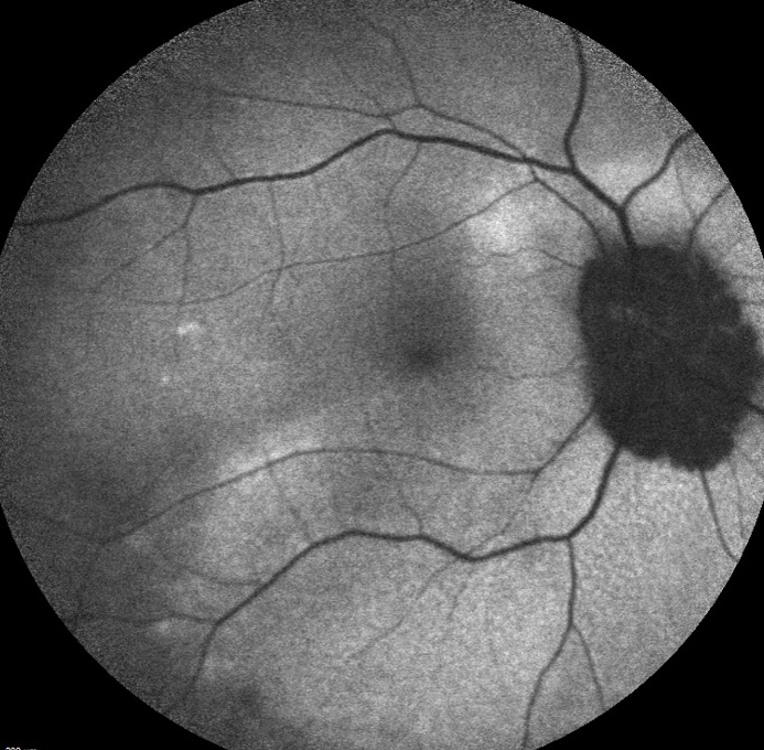
Background: Acute zonal occult outer retinopathy (AZOOR) is a rare autoimmune retinopathy that is challenging to diagnose and treat. It usually presents with subtle fundus changes and severe visual symptoms. Herein, we report a challenging case of AZOOR, emphasizing that multimodal imaging could be valuable in diagnosis and monitoring of treatment response.Case Presentation: A 53-year-old woman presented to the emergency department with a one-week history of subacute, severe, painless vision loss without photopsia in her right eye. Her best-corrected distance visual acuity was 20/800 in the right eye and 20/20 in the left eye. Slit-lamp examination findings were unremarkable, and intraocular pressure was normal in both eyes. Initially, fundus examination findings appeared normal; however, serum levels of inflammatory markers were elevated. Brain and orbital magnetic resonance imaging results were unremarkable. A relative afferent pupillary defect was present in subsequent follow-up examinations at the hospital. The patient initially received a diagnosis of posterior ischemic optic neuropathy secondary to occult giant cell arteritis, underwent steroid treatment, and was evaluated by rheumatology and neurology consultants. Both consultants concurred with the presumed diagnosis. Subsequent multimodal imaging in the ophthalmology clinic revealed a trizonal pattern of fundus autofluorescence. Corresponding to these areas, we noted a loss of the ellipsoid zone on optical coherence tomography, depression on multifocal electroretinogram, and scotoma on visual field testing. Accordingly, the diagnosis of AZOOR was made. The patient was referred back to the rheumatologist for initiation of steroid-sparing treatment, and methotrexate was administered. Five months after the initial presentation, the patient showed significant visual field improvement in both eyes.
Conclusions: Eye care practitioners should consider AZOOR in the differential diagnosis of patients with subacute painless severe unilateral vision loss and unremarkable findings on fundus examination. Multimodal imaging could be valuable in diagnosis and monitoring of treatment response, as observed in the current case. Further studies with larger sample sizes are needed to confirm the value of multimodal imaging and the available management options for AZOOR.
Keywords:
- acute zonal occult outer retinopathy
- white dot syndrome
- giant cell arteritides
- fundus autofluorescence imaging
- electroretinographies
- optical coherence tomography
- visual field test
- Abstract Viewed: 0 times
- Full Text PDF Downloaded: 0 times