Total and outer macular retinal thickness in hydroxychloroquine-treated women with rheumatoid arthritis: a descriptive cross-sectional study
Medical hypothesis, discovery & innovation in optometry,
Vol. 7 No. 3 (2026),
31 October 2026
,
Page 116-122
https://doi.org/10.51329/mehdioptometry252
Abstract
Background: Hydroxychloroquine (HCQ)-associated retinal toxicity may lead to irreversible visual loss, which highlights the importance of accurately characterizing retinal structural measurements in patients receiving long-term treatment. This study aimed to describe device-derived total retinal thickness (TRT) and outer retinal thickness (ORT) measurements obtained using spectral-domain optical coherence tomography (SD-OCT) in women with rheumatoid arthritis (RA) receiving HCQ and healthy female controls.Methods: This descriptive, exploratory cross-sectional study included 64 eye records from 32 women with RA receiving HCQ and 64 eye records from 32 healthy control women. Spectral-domain optical coherence tomography was performed using the REVO FC 130 system (Optopol Technology, Poland). TRT and ORT were summarized for the central, parafoveal, and perifoveal regions of the Early Treatment Diabetic Retinopathy Study grid. The analysis was restricted to descriptive summaries, without inferential comparisons.
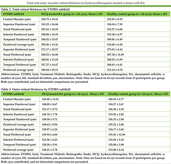
Results: Mean (SD) age was 46.66 (8.90) years in the HCQ-treated RA group and 46.84 (9.13) years in the control group. The corresponding mean (SD) best-corrected visual acuity was 0.04 (0.08) and 0.02 (0.06) logMAR, respectively. In the HCQ-treated RA group, the mean (SD) treatment duration, weight-adjusted daily dose, and cumulative dose were 8.22 (6.85) years, 3.74 (0.97) mg/kg/day, and 1,006.03 (977.80) g, respectively. Mean (SD) central, parafoveal-average, and perifoveal-average TRT measurements were 230.75 (19.41), 305.46 (14.16), and 268.44 (10.11) micrometre, respectively, in the HCQ-treated RA group and 235.81 (8.15), 314.68 (6.10), and 275.14 (6.78) micrometre, respectively, in the control group. The corresponding ORT measurements were 165.48 (13.51), 148.62 (8.56), and 128.21 (5.78) micrometre in the HCQ-treated RA group and 168.42 (6.77), 155.22 (2.68), and 133.00 (4.34) micrometre in the control group.
Conclusions: In this selected cross-sectional sample, descriptive eye-record summaries showed numerically lower mean TRT and ORT measurements in HCQ-treated women with RA than in healthy controls. These observations do not establish population-level differences, HCQ-specific toxicity, selective outer-retinal involvement, temporal progression, or diagnostic biomarker performance. Interpretation is limited by fellow-eye dependence, complete overlap between HCQ exposure and RA status, and the study’s selection and measurement limitations. Future longitudinal studies incorporating an appropriate RA comparison group, accounting for fellow-eye correlation, and using independently defined toxicity outcomes are needed to determine whether ORT has value in detecting early HCQ-related retinal changes.
- hydroxychloroquine
- retinal diseases
- optical coherence tomography
- retina, cross-sectional studies
References
Melles RB, Marmor MF. The risk of toxic retinopathy in patients on long-term hydroxychloroquine therapy. JAMA Ophthalmol. 2014;132(12):1453-1460. doi:10.1001/jamaophthalmol.2014.3459. PMID: 25275721.
Melles RB, Jorge AM, Marmor MF, Zhou B, Conell C, Niu J, McCormick N, Zhang Y, Choi HK. Hydroxychloroquine dose and risk for incident retinopathy: a cohort study. Ann Intern Med. 2023;176(2):166-173. doi:10.7326/M22-2453. PMID: 36645889.
Marmor MF, Ahn SJ, Ehlers JP, Melles RB, Mieler WF, Sarraf D, Yusuf IH; American Academy of Ophthalmology. Special AAO report: recommendations on screening for hydroxychloroquine retinopathy (2025 revision). Ophthalmology. 2026;133(4):439-450. doi:10.1016/j.ophtha.2025.11.001. PMID: 41232611.
Marmor MF, Kellner U, Lai TYY, Melles RB, Mieler WF. Recommendations on screening for chloroquine and hydroxychloroquine retinopathy (2016 revision). Ophthalmology. 2016;123(6):1386-1394. doi:10.1016/j.ophtha.2016.01.058. PMID: 26992838.
Jorge AM, Melles RB, Marmor MF, Zhou B, Zhang Y, Choi HK. Risk Factors for Hydroxychloroquine Retinopathy and Its Subtypes. JAMA Netw Open. 2024 May 1;7(5):e2410677. doi: 10.1001/jamanetworkopen.2024.10677. PMID: 38722628; PMCID: PMC11082687.
Cheong KX, Ong CJT, Chandrasekaran PR, Zhao J, Teo KYC, Mathur R. Review of Retinal Imaging Modalities for Hydroxychloroquine Retinopathy. Diagnostics (Basel). 2023 May 16;13(10):1752. doi: 10.3390/diagnostics13101752. PMID: 37238236; PMCID: PMC10217485.
de Sisternes L, Hu J, Rubin DL, Marmor MF. Localization of damage in progressive hydroxychloroquine retinopathy on and off the drug: inner versus outer retina, parafovea versus peripheral fovea. Invest Ophthalmol Vis Sci. 2015 May;56(5):3415-26. doi: 10.1167/iovs.14-16345. PMID: 26024126; PMCID: PMC4455312.
Mimier-Janczak MK, Kaczmarek D, Proc K, Misiuk-Hoj?o M, Kaczmarek R. Subclinical retinopathy in systemic lupus erythematosus patients - optical coherence tomography study. Reumatologia. 2023;61(3):161-168. doi: 10.5114/reum/166296. Epub 2023 Jun 12. PMID: 37522138; PMCID: PMC10373166.
de Sisternes L, Hu J, Rubin DL, Marmor MF. Analysis of Inner and Outer Retinal Thickness in Patients Using Hydroxychloroquine Prior to Development of Retinopathy. JAMA Ophthalmol. 2016 May 1;134(5):511-519. doi: 10.1001/jamaophthalmol.2016.0155. PMID: 26986043.
Borrelli E, Battista M, Cascavilla ML, Viganò C, Borghesan F, Nicolini N, Clemente L, Sacconi R, Barresi C, Marchese A, Miserocchi E, Modorati G, Bandello F, Querques G. Impact of Structural Changes on Multifocal Electroretinography in Patients With Use of Hydroxychloroquine. Invest Ophthalmol Vis Sci. 2021 Sep 2;62(12):28. doi: 10.1167/iovs.62.12.28. PMID: 34581725; PMCID: PMC8479571.
Tarassoly K, Miraftabi A, Sedaghat A, Parvaresh MM. Analysis of the outer retinal thickness pixel maps for the screening of hydroxychloroquine retinopathy. Int Ophthalmol. 2023 May;43(5):1737-1743. doi: 10.1007/s10792-022-02573-2. Epub 2022 Nov 21. PMID: 36414851.
Salameh N, Abi Doumit C, Jalkh E, Nehme J. Association between hydroxychloroquine intake and damage to the outer nuclear layer in eyes without manifest retinal toxicity. BMC Ophthalmol. 2024;24(1):414. doi:10.1186/s12886-024-03684-3. PMID: 39334015.
Holladay JT. Proper method for calculating average visual acuity. J Refract Surg. 1997;13(4):388-391. doi:10.3928/1081-597X-19970701-16. PMID: 9268940.
Sikorski BL. Ultra-widefield retinal optical coherence tomography (OCT) and angio-OCT using an add-on lens. Diagnostics (Basel). 2025;15(13):1697. doi:10.3390/diagnostics15131697. PMID: 40647696.
Ugwuegbu O, Uchida A, Singh RP, Beven L, Hu M, Kaiser S, Srivastava SK, Ehlers JP. Quantitative assessment of outer retinal layers and ellipsoid zone mapping in hydroxychloroquine retinopathy. Br J Ophthalmol. 2019;103(1):3-7. doi:10.1136/bjophthalmol-2018-312363. PMID: 30190364.
Marmor MF, Chien FY, Johnson MW. Value of red targets and pattern deviation plots in visual field screening for hydroxychloroquine retinopathy. JAMA Ophthalmol. 2013;131(4):476-480. doi:10.1001/jamaophthalmol.2013.1404. PMID: 23710501.
Kim KE, Ryu SJ, Kim YH, Seo Y, Ahn SJ. Visual field examinations using different strategies in Asian patients taking hydroxychloroquine. Sci Rep. 2022 Aug 30;12(1):14778. doi: 10.1038/s41598-022-19048-0. PMID: 36042337; PMCID: PMC9427842.
Birt CM, Shin DH, Samudrala V, Hughes BA, Kim C, Lee D. Analysis of reliability indices from Humphrey visual field tests in an urban glaucoma population. Ophthalmology. 1997;104(7):1126-1130. doi:10.1016/S0161-6420(97)30173-0. PMID: 9224465.
Kim KE, Kim JH, Kim YH, Ahn SJ. Clock-hour topography and extent of outer retinal damage in hydroxychloroquine retinopathy. Sci Rep. 2022 Jul 12;12(1):11809. doi: 10.1038/s41598-022-15217-3. PMID: 35821509; PMCID: PMC9276819.
Casado A, López-de-Eguileta A, Fonseca S, Muñoz P, Demetrio R, Gordo-Vega MA, Cerveró A. Outer Nuclear Layer Damage for Detection of Early Retinal Toxicity of Hydroxychloroquine. Biomedicines. 2020 Mar 4;8(3):54. doi: 10.3390/biomedicines8030054. PMID: 32143497; PMCID: PMC7148510.
Ferreira A, Anjos R, José-Vieira R, Afonso M, Abreu AC, Monteiro S, Macedo M, Andrade JP, Furtado MJ, Lume M. Application of optical coherence tomography angiography for microvascular changes in patients treated with hydroxychloroquine: a systematic review and meta-analysis. Graefes Arch Clin Exp Ophthalmol. 2023 Aug;261(8):2221-2233. doi: 10.1007/s00417-023-06023-2. Epub 2023 Mar 8. PMID: 36884062; PMCID: PMC9993382.
Yusuf IH, Charbel Issa P, Ahn SJ. Hydroxychloroquine-induced retinal toxicity. Front Pharmacol. 2023;14:1196783. doi:10.3389/fphar.2023.1196783. PMID: 37324471.
Mahon GJ, Anderson HR, Gardiner TA, McFarlane S, Archer DB, Stitt AW. Chloroquine causes lysosomal dysfunction in neural retina and RPE: implications for retinopathy. Curr Eye Res. 2004 Apr;28(4):277-84. doi: 10.1076/ceyr.28.4.277.27835. PMID: 15259297.
Uslu H, Gurler B, Yildirim A, Tatar MG, Kantarci FA, Goker H, Pehlevan HS, Colak HN. Effect of Hydroxychloroquine on the Retinal Layers: A Quantitative Evaluation with Spectral-Domain Optical Coherence Tomography. J Ophthalmol. 2016;2016:8643174. doi: 10.1155/2016/8643174. Epub 2016 Aug 30. PMID: 27656292; PMCID: PMC5021475.
Montesel A, Sacconi R, La Rubia P, Querques G. Impact of cumulative dose of hydroxychloroquine on retinal structures. Graefes Arch Clin Exp Ophthalmol. 2025 Jan;263(1):235-237. doi: 10.1007/s00417-024-06602-x. Epub 2024 Aug 19. PMID: 39160441.
Zhao H, Pan M, Liu Y, Cheng F, Shuai Z. Evaluation of early retinal changes in patients on long-term hydroxychloroquine using optical coherence tomography angiography. Ther Adv Drug Saf. 2024 Jan 27;15:20420986231225851. doi: 10.1177/20420986231225851. PMID: 38300766; PMCID: PMC10823852.
Pasadhika S, Fishman GA, Choi D, Shahidi M. Selective thinning of the perifoveal inner retina as an early sign of hydroxychloroquine retinal toxicity. Eye (Lond). 2010 May;24(5):756-62; quiz 763. doi: 10.1038/eye.2010.21. Epub 2010 Apr 16. PMID: 20395978; PMCID: PMC3711255.
Jung SH, Park YH, Park YG. Subclinical Detection of Hydroxychloroquine-Induced Retinopathy in Patients with Systemic Lupus Erythematous Using Multifocal Electroretinography and Optical Coherence Tomography. J Clin Med. 2024 Dec 16;13(24):7663. doi: 10.3390/jcm13247663. PMID: 39768585; PMCID: PMC11678038.
- Abstract Viewed: 0 times
- Full Text PDF Downloaded: 0 times