Presumed herpes simplex virus keratouveitis in a 2-year-old child: a case report
Medical hypothesis discovery and innovation in ophthalmology,
Vol. 15 No. 3 (2026),
12 October 2026
,
Page 238-251
https://doi.org/10.51329/mehdiophthal1553
Abstract
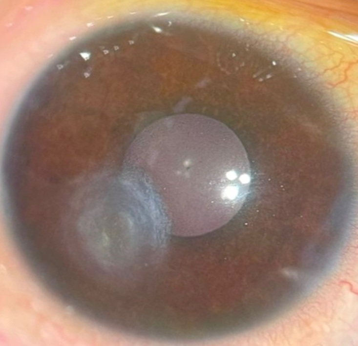
Background: Ocular herpes simplex virus (HSV) disease is uncommon in very young children and may be difficult to recognize because history and examination are limited and early manifestations can resemble bacterial conjunctivitis. Pediatric disease may involve multiple corneal layers, extend into the anterior chamber, recur, and threaten visual development. We describe presumed HSV keratouveitis in a 2-year-old child.Case Presentation: A 2-year-old girl presented with a two-month history of right-eye pain, photophobia, and tearing after an episode diagnosed elsewhere as HSV blepharoconjunctivitis. Previous topical treatments, including antibiotics, had produced little improvement. Examination revealed right eyelid edema, conjunctival congestion, and an inferior geographic epithelial defect involving one-third of the cornea; the anterior chamber was quiet. Suspected HSV epithelial keratitis was treated with topical acyclovir 3% ointment, moxifloxacin, and lubrication. Although the epithelium healed, stromal edema, infiltration, and endotheliitis developed. Topical prednisolone acetate and therapeutic-dose oral acyclovir were commenced after normal renal function was confirmed. Serological testing showed reactive cytomegalovirus (CMV) IgG and IgM; Epstein-Barr virus IgG and IgM, HSV-1 IgG, HSV-2 IgG, rubella IgM, and Toxoplasma IgG and IgM were non-reactive. Initial improvement was followed by acute recurrence one week after transition to prophylactic acyclovir. Examination under anesthesia revealed a recurrent 3-mm epithelial defect, diffuse stromal edema and infiltration, 2+ anterior chamber cells, and right-eye intraocular pressure of 29 mmHg; no keratic precipitates, iris atrophy, synechiae, or posterior-segment abnormalities were identified. Ocular-surface HSV-1/HSV-2 polymerase chain reaction (PCR), obtained after four weeks of topical and three weeks of systemic acyclovir, was negative, and bacterial cultures showed no growth. Ocular CMV and varicella-zoster virus testing was not performed, and the parents declined aqueous-humor sampling. The child received intravenous acyclovir (10 mg/kg every 8 hours) and methylprednisolone (1 mg/kg/day) for five days, with topical anti-inflammatory, cycloplegic, pressure-lowering, lubricating, and initially empirical antibacterial treatment. At discharge, the eye was quiet, intraocular pressure had normalized, and a 4-mm paracentral stromal opacity remained. At six months there was no recurrence; intraocular pressure was 15 mmHg, refraction was stable, and no fixation preference or ocular misalignment was observed.
Conclusions: The sequential unilateral epithelial, stromal, endothelial, and anterior uveal involvement favored HSV, but absent aqueous-humor testing and negative post-treatment surface PCR precluded virological confirmation. This case supports longitudinal assessment of persistent unilateral corneal inflammation in young children and surveillance for recurrence, corneal sequelae, and impaired visual development.
- Abstract Viewed: 0 times
- Full Text PDF Downloaded: 0 times


