Macular Inner Retinal Layer Thinning in Diabetic Patients without Retinopathy Measured by Spectral Domain Optical Coherence Tomography
Medical hypothesis discovery and innovation in ophthalmology,
Vol. 7 No. 3 (2018),
1 September 2018
,
Page 133-139
Abstract
The aim of this study was to use Spectral Domain-Optical Coherence Tomography (SD-OCT) to measure the thickness of the Macular Inner Retinal Layer (MIRL) and compare the results between diabetic patients with no signs of retinopathy and healthy subjects. Overall, 47 type 2 diabetic patients without clinical signs of retinopathy were prospectively analyzed along with 36 healthy subjects. This study excluded patients with other systemic or ocular diseases. All patients had their MIRL thickness measured by RTVue-100 SD-OCT (7x7 mm macular grid). The MIRL thickness is provided by the ganglion cell complex scan (comprised of the retinal nerve fiber, ganglion cell, and inner plexiform layers). Only one eye was randomly selected if both were eligible for analysis. Mean age was similar between the two groups (diabetic patients: 57.3 ± 10.6 and control subjects: 60.2 ± 12.2 years) (P = 0.19). No significant differences regarding optic disc area and cup-to-disc ratio was observed in the comparison of the two groups (P ≥ 0.38 for both comparisons). In patients with diabetes, the average MIRL was significantly thinner when compared to controls (91.6 versus 96.2 micrometer (µm); P = 0.02). Regional analysis revealed superior and inferior MIRL to be significantly thinner in patients with diabetes than the controls (P ≤ 0.04). The juxtafoveal area was compromised (thinned) in 70% of diabetic eyes, classified as abnormal (P < 1%; compared to the device’s normative database). In conclusion, patients with type 2 diabetes without clinical evidence of retinopathy had lower MIRL average values when compared to the control group. This can be explained by the ischemia and retinal tissue injury caused by diabetes even in early stages of diabetic retinopathy, which can affect MIRL thickness. Possible implications of these findings on diagnosis and treatment of diabetic retinopathy requires further investigation.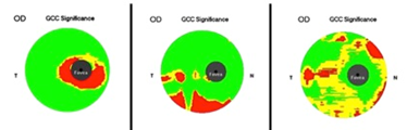
References
Frank RN. On the pathogenesis of diabetic retinopathy. Ophthalmology. 1984;91(6):626-34. pmid: 6205341
Lieth E, Gardner TW, Barber AJ, Antonetti DA, Penn State Retina Research G. Retinal neurodegeneration: early pathology in diabetes. Clin Exp Ophthalmol. 2000;28(1):3-8. pmid: 11345341
Chihara E, Matsuoka T, Ogura Y, Matsumura M. Retinal nerve fiber layer defect as an early manifestation of diabetic retinopathy. Ophthalmology. 1993;100(8):1147-51. pmid: 8341494
Lopes de Faria JM, Russ H, Costa VP. Retinal nerve fibre layer loss in patients with type 1 diabetes mellitus without retinopathy. Br J Ophthalmol. 2002;86(7):725-8. pmid: 12084737
Kim K, Kim ES, Yu SY. Longitudinal relationship between retinal diabetic neurodegeneration and progression of diabetic retinopathy in patients with type 2 diabetes. Am J Ophthalmol. 2018. doi: 10.1016/j.ajo.2018.08.053 pmid: 30195892
Barber AJ, Lieth E, Khin SA, Antonetti DA, Buchanan AG, Gardner TW. Neural apoptosis in the retina during experimental and human diabetes. Early onset and effect of insulin. J Clin Invest. 1998;102(4):783-91. doi: 10.1172/JCI2425 pmid: 9710447
Gardner TW, Antonetti DA, Barber AJ, LaNoue KF, Levison SW. Diabetic retinopathy: more than meets the eye. Surv Ophthalmol. 2002;47 Suppl 2:S253-62. pmid: 12507627
Huang D, Swanson EA, Lin CP, Schuman JS, Stinson WG, Chang W, et al. Optical coherence tomography. Science. 1991;254(5035):1178-81. pmid: 1957169
Hee MR, Izatt JA, Swanson EA, Huang D, Schuman JS, Lin CP, et al. Optical coherence tomography of the human retina. Arch Ophthalmol. 1995;113(3):325-32. pmid: 7887846
Puliafito CA, Hee MR, Lin CP, Reichel E, Schuman JS, Duker JS, et al. Imaging of macular diseases with optical coherence tomography. Ophthalmology. 1995;102(2):217-29. pmid: 7862410
Drexler W. Ultrahigh-resolution optical coherence tomography. J Biomed Opt. 2004;9(1):47-74. doi: 10.1117/1.1629679 pmid: 14715057
Nassif N, Cense B, Park BH, Yun SH, Chen TC, Bouma BE, et al. In vivo human retinal imaging by ultrahigh-speed spectral domain optical coherence tomography. Opt Lett. 2004;29(5):480-2. pmid: 15005199
Podoleanu AG, Rosen RB. Combinations of techniques in imaging the retina with high resolution. Prog Retin Eye Res. 2008;27(4):464-99. doi: 10.1016/j.preteyeres .2008.03.002 pmid: 18495519
Srinivasan VJ, Wojtkowski M, Witkin AJ, Duker JS, Ko TH, Carvalho M, et al. High-definition and 3-dimensional imaging of macular pathologies with high-speed ultrahigh-resolution optical coherence tomography. Ophthalmology. 2006;113(11):2054 e1-14. doi: 10.1016/j.ophtha.2006.05.046 pmid: 17074565
Sakamoto A, Hangai M, Yoshimura N. Spectral-domain optical coherence tomography with multiple B-scan averaging for enhanced imaging of retinal diseases. Ophthalmology. 2008;115(6):1071-8 e7. doi: 10.1016/j.ophtha.2007.09.001 pmid: 18061270
Jittpoonkuson T, Garcia PM, Rosen RB. Correlation between fluorescein angiography and spectral-domain optical coherence tomography in the diagnosis of cystoid macular edema. Br J Ophthalmol. 2010;94(9):1197-200. doi: 10.1136/bjo.2009.170589 pmid: 19965832
Gama R, Relha C, Gomes Costa J, Eiro N. Measurement of the Inner Retinal Layers of Megalopapilla by Optical Coherence Tomography. Med Hypothesis Discov Innov Ophthalmol. 2017;6(3):82-8. pmid: 29392147
Tan O, Chopra V, Lu AT, Schuman JS, Ishikawa H, Wollstein G, et al. Detection of macular ganglion cell loss in glaucoma by Fourier-domain optical coherence tomography. Ophthalmology. 2009;116(12):2305-14 e1-2. doi: 10.1016/j.ophtha.2009.05.025 pmid: 19744726
Prata TS, Dorairaj S, Trancoso L, Kanadani FN, Biteli LG, Furlanetto R, et al. Eyes with large disc cupping and normal intraocular pressure: using optical coherence tomography to discriminate those with and without glaucoma. Med Hypothesis Discov Innov Ophthalmol. 2014;3(3):91-8. pmid: 25741525
Tekin K, Inanc M, Kurnaz E, Bayramoglu E, Aydemir E, Koc M, et al. Quantitative evaluation of early retinal changes in children with type 1 diabetes mellitus without retinopathy. Clin Exp Optom. 2018;101(5):680-5. doi: 10.1111/cxo.12667 pmid: 29488254
Pekel E, Altincik SA, Pekel G. Evaluation of optic disc, retinal nerve fiber and macular ganglion cell layers in pediatric diabetes. Int Ophthalmol. 2018;38(5):1955-61. doi: 10.1007/s10792-017-0683-3 pmid: 28780619
Kim K, Kim ES, Yu SY. Optical coherence tomography angiography analysis of foveal microvascular changes and inner retinal layer thinning in patients with diabetes. Br J Ophthalmol. 2018;102(9):1226-31. doi: 10.1136/bjophthalmol-2017-311149 pmid: 29259019
Sahin M, Sahin A, Kilinc F, Karaalp U, Yuksel H, Ozkurt ZG, et al. Early detection of macular and peripapillary changes with spectralis optical coherence tomography in patients with prediabetes. Arch Physiol Biochem. 2018;124(1):75-9. doi: 10.1080/13813455.2017.13614 50 pmid: 28780883
Pekel E, Tufaner G, Kaya H, Kasikci A, Deda G, Pekel G. Assessment of optic disc and ganglion cell layer in diabetes mellitus type 2. Medicine (Baltimore). 2017;96(29):e7556. doi: 10.1097/MD.000000000000 7556 pmid: 28723781
Li ST, Wang XN, Du XH, Wu Q. Comparison of spectral-domain optical coherence tomography for intra-retinal layers thickness measurements between healthy and diabetic eyes among Chinese adults. PLoS One. 2017;12(5):e0177515. doi: 10.1371/journal.pone.017 7515 pmid: 28493982
Cabrera DeBuc D, Somfai GM. Early detection of retinal thickness changes in diabetes using Optical Coherence Tomography. Med Sci Monit. 2010;16(3):MT15-21. pmid: 20190693
Hegazy AI, Zedan RH, Macky TA, Esmat SM. Retinal ganglion cell complex changes using spectral domain optical coherence tomography in diabetic patients without retinopathy. Int J Ophthalmol. 2017;10(3):427-33. doi: 10.18240/ijo.2017.03.16 pmid: 28393035
Choi JA, Kim HW, Kwon JW, Shim YS, Jee DH, Yun JS, et al. Early inner retinal thinning and cardiovascular autonomic dysfunction in type 2 diabetes. PLoS One. 2017;12(3):e0174377. doi: 10.1371/journal.pone.017 4377 pmid: 28334035
Wilkinson CP, Ferris FL, 3rd, Klein RE, Lee PP, Agardh CD, Davis M, et al. Proposed international clinical diabetic retinopathy and diabetic macular edema disease severity scales. Ophthalmology. 2003;110(9):1677-82. doi: 10.1016/S0161-6420(03)00 475-5 pmid: 13129861
Seong M, Sung KR, Choi EH, Kang SY, Cho JW, Um TW, et al. Macular and peripapillary retinal nerve fiber layer measurements by spectral domain optical coherence tomography in normal-tension glaucoma. Invest Ophthalmol Vis Sci. 2010;51(3):1446-52. doi: 10.1167/iovs.09-4258 pmid: 19834029
Tan O, Li G, Lu AT, Varma R, Huang D, Advanced Imaging for Glaucoma Study G. Mapping of macular substructures with optical coherence tomography for glaucoma diagnosis. Ophthalmology. 2008;115(6):949-56. doi: 10.1016/j.ophtha.2007.08.0 11 pmid: 17981334
Abu-El-Asrar AM, Dralands L, Missotten L, Al-Jadaan IA, Geboes K. Expression of apoptosis markers in the retinas of human subjects with diabetes. Invest Ophthalmol Vis Sci. 2004;45(8):2760-6. doi: 10.1167/iovs.03-1392 pmid: 15277502
Chihara E, Zhang S. [Analysis of diabetic optic neuropathy with a topographic laser scanning system]. Nippon Ganka Gakkai Zasshi. 1998;102(7):431-5. pmid: 9720364
Simonsen SE. The value of the oscillatory potential in selecting juvenile diabetics at risk of developing proliferative retinopathy. Acta Ophthalmol (Copenh). 1980;58(6):865-78. pmid: 7331773
Juen S, Kieselbach GF. Electrophysiological changes in juvenile diabetics without retinopathy. Arch Ophthalmol. 1990;108(3):372-5. pmid: 2310337
van Dijk HW, Verbraak FD, Kok PH, Garvin MK, Sonka M, Lee K, et al. Decreased retinal ganglion cell layer thickness in patients with type 1 diabetes. Invest Ophthalmol Vis Sci. 2010;51(7):3660-5. doi: 10.1167/iovs.09-5041 pmid: 20130282
Tretiach M, Madigan MC, Wen L, Gillies MC. Effect of Muller cell co-culture on in vitro permeability of bovine retinal vascular endothelium in normoxic and hypoxic conditions. Neurosci Lett. 2005;378(3):160-5. doi: 10.1016/j.neulet.2004.12.026 pmid: 15781151
- Abstract Viewed: 1253 times
- Full Text PDF Downloaded: 1067 times
