The Correlation between Corneal Topographic Indices and Corneal High Order Aberrations in Keratoconus
Medical hypothesis discovery and innovation in ophthalmology,
Vol. 8 No. 1 (2019),
10 March 2019
,
Page 1-6
Abstract
This study was performed to investigate aberrometric changes of Keratoconus (KC) and its correlation with corneal topographic indices. In a cross sectional study, the researchers included 170 eyes of 138 candidates that were seeking corneal refractive surgery in Sohag refractive center, Sohag, Egypt and had been diagnosed as clinical KC. Patients were divided to mild, moderate, and severe KC. All eyes included in this study were subjected to corneal tomographic evaluation. Corneal aberrometry data was collected from the Sirius topography (Sirius, Costruzione Strumenti Oftalmici, Italy) over a 5-mm diameter. The collected data included Zernike coefficients for corneal aberrations, including total Root Mean Square (RMS), RMS Spherical Aberration (SA), RMS Coma, and RMS astigmatism. The study population was divided to mild, moderate, and severe KC. Mild KC cases included 58 eyes of 46 patients, moderate KC were 64 eyes of 52 patients, and severe KC were 48 eyes of 40 patients. Root mean square total was statistically significant in all groups with a higher p value in moderate KC (P = 0.001) and also was statistically significant when compared in the three groups altogether (P = 0.0001). Coma aberration was statistically significant in mild and moderate KC and when compared between the three groups (P = 0.0001). Root mean square Trefoil aberration was statistically significant only in moderate KC yet was statistically significant when compared in all groups (P = 0.0001). Root mean square astigmatism was statistically significant in mild KC only and when compared in the three groups altogether (P = 0.0001). Spherical aberration was also statistically significant in moderate and severe KC with a P value of < 0.0001 and 0.001, respectively. There was a positive correlation between posterior elevation and RMS values in mild and moderate KC while there was negative or very weak positive correlation in severe KC. There were negative correlations between the thinnest location and RMS values in nearly all variables in the three groups except weak positive correlation with RMS astigmatism in mild KC and with RMS total and coma aberration in severe KC .In conclusion corneal high order aberrations measured by the Sirius topography system had low to moderate correlation with corneal topographic indices provided by the same device in different grades of KC.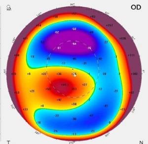
References
Rabinowitz YS. Keratoconus. Surv Ophthalmol 1998;42(4):297-319. doi: 10.1016/s0039-6257(97)001 19-7
Sugar J, Macsai MS. What causes keratoconus? Cornea. 2012;31(6):716-9. doi: 10.1097/ICO.0b013e31 823f8c72 pmid: 22406940
Gatzioufas Z, Panos GD, Hamada S. Keratoconus: is it a Non-inflammatory Disease? Med Hypothesis Discov Innov Ophthalmol. 2017;6(1):1-2. pmid: 28428967
Applegate RA, Hilmantel G, Howland HC, Tu EY, Starck T, Zayac EJ. Corneal first surface optical aberrations and visual performance. J Refract Surg. 2000;16(5):507-14. pmid: 11019864
Maeda N, Klyce SD, Smolek MK, Thompson HW. Automated keratoconus screening with corneal topography analysis. Invest Ophthalmol Vis Sci. 1994;35(6):2749-57. pmid: 8188468
Nilforoushan MR, Speaker M, Marmor M, Abramson J, Tullo W, Morschauser D, et al. Comparative evaluation of refractive surgery candidates with Placido topography, Orbscan II, Pentacam, and wavefront analysis. J Cataract Refract Surg. 2008;34(4):623-31. doi: 10.1016/j.jcrs.2007.11.054 pmid: 18361985
Ucakhan OO, Cetinkor V, Ozkan M, Kanpolat A. Evaluation of Scheimpflug imaging parameters in subclinical keratoconus, keratoconus, and normal eyes. J Cataract Refract Surg. 2011;37(6):1116-24. doi: 10.1016/j.jcrs.2010.12.049 pmid: 21596255
de Sanctis U, Loiacono C, Richiardi L, Turco D, Mutani B, Grignolo FM. Sensitivity and specificity of posterior corneal elevation measured by Pentacam in discriminating keratoconus/subclinical keratoconus. Ophthalmology. 2008;115(9):1534-9. doi: 10.1016/j.o phtha.2008.02.020 pmid: 18405974
Jafri B, Li X, Yang H, Rabinowitz YS. Higher order wavefront aberrations and topography in early and suspected keratoconus. J Refract Surg. 2007;23(8):774-81. pmid: 17985796
Pantanelli S, MacRae S, Jeong TM, Yoon G. Characterizing the wave aberration in eyes with keratoconus or penetrating keratoplasty using a high-dynamic range wavefront sensor. Ophthalmology. 2007;114(11):2013-21. doi: 10.1016/j.ophtha.2007. 01.008 pmid: 17553566
Gobbe M, Guillon M. Corneal wavefront aberration measurements to detect keratoconus patients. Cont Lens Anterior Eye. 2005;28(2):57-66. doi: 10.1016/j.clae.2004.12.001 pmid: 16318836
Alio JL, Shabayek MH. Corneal higher order aberrations: a method to grade keratoconus. J Refract Surg. 2006;22(6):539-45. pmid: 16805116
Prakash G, Srivastava D, Suhail M, Bacero R. Assessment of bilateral pupillary centroid characteristics at varying illuminations and post-photopic flash response using an automated pupillometer. Clin Exp Optom. 2016;99(6):535-43. doi: 10.1111/cxo.12409 pmid: 27432474
Lombardo M, Lombardo G. Wave aberration of human eyes and new descriptors of image optical quality and visual performance. J Cataract Refract Surg. 2010;36(2):313-31. doi: 10.1016/j.jcrs.2009.09.026 pmid: 20152616
Mahmoud AM, Nunez MX, Blanco C, Koch DD, Wang L, Weikert MP, et al. Expanding the cone location and magnitude index to include corneal thickness and posterior surface information for the detection of keratoconus. Am J Ophthalmol. 2013;156(6):1102-11. doi: 10.1016/j.ajo.2013.07.018 pmid: 24075426
Saad A, Gatinel D. Evaluation of total and corneal wavefront high order aberrations for the detection of forme fruste keratoconus. Invest Ophthalmol Vis Sci. 2012;53(6):2978-92. doi: 10.1167/iovs.11-8803 pmid: 22427590
Hallahan KM, Sinha Roy A, Ambrosio R, Jr., Salomao M, Dupps WJ, Jr. Discriminant value of custom ocular response analyzer waveform derivatives in keratoconus. Ophthalmology. 2014;121(2):459-68. doi: 10.1016/j.ophtha.2013.09.013 pmid: 24289916
Colak HN, Kantarci FA, Yildirim A, Tatar MG, Goker H, Uslu H, et al. Comparison of corneal topographic measurements and high order aberrations in keratoconus and normal eyes. Cont Lens Anterior Eye. 2016;39(5):380-4. doi: 10.1016/j.clae.2016.06.005 pmid: 27395753
Maeda N, Fujikado T, Kuroda T, Mihashi T, Hirohara Y, Nishida K, et al. Wavefront aberrations measured with Hartmann-Shack sensor in patients with keratoconus. Ophthalmology. 2002;109(11):1996-2003. doi: 10.1016/s0161-6420(02)01279-4 pmid: 12414405
Delgado S, Velazco J, Delgado Pelayo RM, Ruiz-Quintero N. Correlation of higher order aberrations in the anterior corneal surface and degree of keratoconus measured with a Scheimpflug camera. Arch Soc Esp Oftalmol. 2016;91(7):316-9. doi: 10.1016/j.oftal.2016.01.014 pmid: 26907199
Nakagawa T, Maeda N, Kosaki R, Hori Y, Inoue T, Saika M, et al. Higher-order aberrations due to the posterior corneal surface in patients with keratoconus. Invest Ophthalmol Vis Sci. 2009;50(6):2660-5. doi: 10.1167/iovs.08-2754 pmid: 19029032
Schlegel Z, Lteif Y, Bains HS, Gatinel D. Total, corneal, and internal ocular optical aberrations in patients with keratoconus. J Refract Surg. 2009;25(10 Suppl):S951-7. doi: 10.3928/1081597X-20090915-10 pmid: 19848377
Alio JL, Pinero DP, Aleson A, Teus MA, Barraquer RI, Murta J, et al. Keratoconus-integrated characterization considering anterior corneal aberrations, internal astigmatism, and corneal biomechanics. J Cataract Refract Surg. 2011;37(3):552-68. doi: 10.1016/j.jcrs. 2010.10.046 pmid: 21333878
Ramin S, Sangin Abadi A, Doroodgar F, Esmaeili M, Niazi F, Niazi S, et al. Comparison of Visual, Refractive and Aberration Measurements of INTACS versus Toric ICL Lens Implantation; A Four-year Follow-up. Med Hypothesis Discov Innov Ophthalmol. 2018;7(1):32-9. pmid: 29644243
- Abstract Viewed: 1251 times
- Full Text PDF Downloaded: 910 times
