Intrafamilial Phenotype Variability in Two Male Siblings, With X-linked Juvenile Retinoschisis and Dorzolamide Treatment Effect in the Natural History of the Disease
Medical hypothesis discovery and innovation in ophthalmology,
Vol. 8 No. 1 (2019),
10 March 2019
,
Page 11-15
Abstract
To investigate how genotype is related to phenotype and document correlations of genotype-phenotype with response of topical administration of dorzolamide in siblings affected with X-linked juvenile retinoschisis (XLRS). We performed a retrospective study on two male siblings (four eyes) with XLRS, who were treated with topical installation of dorzolamide. Clinical diagnosis was supported with familial genetic analysis with bi-directional Sanger sequencing of RS1 pathogenic variant. Optical coherence tomography (OCT), fundus fluorescein angiography (FFA), ultrasound scan (U/S) and electroretinogram (ERG) were used in the evaluation. Central macular thickness (CMT) and best corrected visual acuity (BCVA) were recorded monthly for eighteen months. We performed genetic analysis in their family for mutations in the gene that encodes the protein retinoschisin, responsible for retinoschisis (RS1). It was proved that phenotype variability might be related to the same pathogenic variant. While there was an improvement in BCVA and OCT central macular thickness in the patient with the mild form of disease, the visual acuity and the OCT scans of the patient with severe form of disease did not improve. Intrafamilial phenotypic variability between individuals sharing identical pathogenic variant was documented. Both our patients had a pathogenic variant in a hemizygous state at a genomic location in exon 6 of the RS1 gene; Frameshift mutation that is likely to cause protein truncation was identified which is suggested to result in greater clinical severity. Consequently, it was found that response to dorzolamide is correlated to phenotypic severity.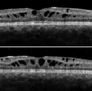
References
Functional Implications of the Spectrum of Mutations Found in 234 Cases With X-linked Juvenile Retinoschisis (XLRS). Hum Mol Gen. 1998;7(7):1185-92. doi: 10.1093/hmg/7.7.1185
Sauer CG, Gehrig A, Warneke-Wittstock R, Marquardt A, Ewing CC, Gibson A, et al. Positional cloning of the gene associated with X-linked juvenile retinoschisis. Nat Genet. 1997;17(2):164-70. doi: 10.1038/ng1097-164 pmid: 9326935
Apushkin MA, Fishman GA, Rajagopalan AS. Fundus findings and longitudinal study of visual acuity loss in patients with X-linked retinoschisis. Retina. 2005;25(5):612-8. pmid: 16077359
Kim LS, Seiple W, Fishman GA, Szlyk JP. Multifocal ERG findings in carriers of X-linked retinoschisis. Doc Ophthalmol. 2007;114(1):21-6. doi: 10.1007/s10633-006-9034-9 pmid: 17180613
Sieving P. Juvenile retinoschisis. In: Traboulsi E, editor. Genetic Diseases of the Eye. New York, NY: Oxford University Press; 1998. p. 347-55.
Nakamura M, Ito S, Terasaki H, Miyake Y. Japanese X-linked juvenile retinoschisis: conflict of phenotype and genotype with novel mutations in the XLRS1 gene. Arch Ophthalmol. 2001;119(10):1553-4. pmid: 11594966
George ND, Yates JR, Moore AT. Clinical features in affected males with X-linked retinoschisis. Arch Ophthalmol. 1996;114(3):274-80. pmid: 8600886
Sieving PA, Bingham EL, Kemp J, Richards J, Hiriyanna K. Juvenile X-linked retinoschisis from XLRS1 Arg213Trp mutation with preservation of the electroretinogram scotopic b-wave. American Journal of Ophthalmology. 1999;128(2):179-84. doi: 10.1016/s0002-9394(99)00144-0
Azzolini C, Pierro L, Codenotti M, Brancato R. Oct Images and Surgery of Juvenile Macular Retinoschisis. European Journal of Ophthalmology. 2018;7(2):196-200. doi: 10.1177/112067219700700214
Fishman GA. Acetazolamide for Treatment of Chronic Macular Edema in Retinitis Pigmentosa. Archives of Ophthalmology. 1989;107(10):1445. doi: 10.1001/archopht.1989.01070020519031
Grover S, Fishman GA, Fiscella RG, Adelman AE. Efficacy of dorzolamide hydrochloride in the management of chronic cystoid macular edema in patients with retinitis pigmentosa. Retina. 1997;17(3):222-31. pmid: 9196934
Genead MA, Fishman GA, Walia S. Efficacy of sustained topical dorzolamide therapy for cystic macular lesions in patients with X-linked retinoschisis. Arch Ophthalmol. 2010;128(2):190-7. doi: 10.1001/archophthalmol.2009.398 pmid: 20142541
Ajlan RS, Hammamji KS. Stellate Nonhereditary Idiopathic Foveomacular Retinoschisis: Response to Topical Dorzolamide Therapy. Retin Cases Brief Rep. 2017. doi: 10.1097/ICB.0000000000000599 pmid: 28557864
Wu WW, Wong JP, Kast J, Molday RS. RS1, a discoidin domain-containing retinal cell adhesion protein associated with X-linked retinoschisis, exists as a novel disulfide-linked octamer. J Biol Chem. 2005;280(11):10721-30. doi: 10.1074/jbc.M413117200 pmid: 15644328
Coussa RG, Kapusta MA. Treatment of cystic cavities in X-linked juvenile retinoschisis: The first sequential cross-over treatment regimen with dorzolamide. Am J Ophthalmol Case Rep. 2017;8:1-3. doi: 10.1016/j.ajoc.2017.07.008 pmid: 29260104
Kim DY, Mukai S. X-linked juvenile retinoschisis (XLRS): a review of genotype-phenotype relationships. Semin Ophthalmol. 2013;28(5-6):392-6. doi: 10.3109/08820538.2013.825299 pmid: 24138048
- Abstract Viewed: 1228 times
- Full Text PDF Downloaded: 908 times
