Diagnosis and Management of Pseudoguttata: A Literature Review
Medical hypothesis discovery and innovation in ophthalmology,
Vol. 8 No. 3 (2019),
20 September 2019
,
Page 156-162
Abstract
Corneal pseudoguttata (PG), also known as pseudoguttae or secondary guttata, is a transient, reversible endothelial edema commonly associated with anterior segment pathology. While considered rare, PG presents on slit-lamp examination more commonly than originally thought. We have clinically observed PG after refractive surgeries, in association with infectious keratitis, and following medication use. PG presents as dark lesions on slit-lamp exam with specular illumination, similar to primary corneal guttata. PG is distinct from guttata because PG resolves over time and does not involve Descemet’s membrane. Other ocular findings that may be confused with guttata include endothelial blebs (EB) and endothelial denudation (ED). EB are possibly a type of PG that present after contact lens use or hypoxia. ED is a distinct entity that is characterized by loss of endothelial cells without involvement of Descemet’s membrane. Confocal microscopy may be useful in differentiating these four endothelial lesions, with differences in border definition and the presence of hyperreflective areas two main distinctions. PG presents as a hyporeflective, elevated shape without clear borders on confocal microscopy. PG, EB, and ED can resolve with time without the need for surgical intervention, unlike corneal guttata. Treatment of the underlying condition will lead to resolution of both PG and EB.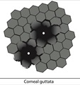
References
Zavala J, Lopez Jaime GR, Rodriguez Barrientos CA, Valdez-Garcia J. Corneal endothelium: developmental strategies for regeneration. Eye (Lond). 2013;27:579-88. doi: 10.1038/eye.2013.15 pmid: 23470788
Eghrari AO, Gottsch JD. Fuchs' corneal dystrophy. Expert Rev Ophthalmol. 2010;5:147-59. doi: 10.1586/eop.10.8 pmid: 20625449
Krachmer JH, Schnitzer JI, Fratkin J. Cornea pseudoguttata: a clinical and histopathologic description of endothelial cell edema. Arch Ophthalmol. 1981;99:1377-81. doi: 10.1001/archopht.1981.03930020251007 pmid: 7259610
Doughty MJ, Jonuscheit S, Button NF. Central corneal thickness and intraocular pressure measures in human corneas with endothelial guttata: an observational quality control study. Clin Exp Optom. 2011;94:425-32. doi: 10.1111/j.1444-0938.2011.00584.x pmid: 21777286
Vogt A. Weitere Ergebnisse der Spaltlampenmikroskopie des vordern Bulbusabschnittes. Albrecht von Græfes Archiv für Ophthalmologie. 1921;106:63-103.
Rett D. BLOG: Guttae or guttata? Some thoughts on Fuchs’. [cited 2019 June 3]. Available from: https://www.healio.com/optometry/cornea-external-disease/news/blogs/%7Bc9bcca64-1c9e-4787-924a-12dd0c779837%7D/doug-rett-od-faao/blog-guttae-or-guttata-some-thoughts-on-fuchs.
Lietman T, Lee J, Costanza S. Those excrescences on Descemet's membrane. Br J Ophthalmol. 2003;87:515-6. doi: 10.1136/bjo.87.4.515-a pmid: 12642334
Dore S. Two Cases of Morphoea Guttata. 1917 [cited 2019 May 29]. Available from: https://journals.sagepub.com/doi/pdf/10.1177/003591571801100217.
Wolter JR, Larson BF. Pathology of Cornea Guttata*. American Journal of Ophthalmology. 1959;48:161-9. doi: 10.1016/0002-9394(59)90255-7
Zantos SG, Holden BA. Transient Endothelial Changes Soon After Wearing Soft Contact Lenses. Optometry and Vision Science. 1977;54:856-8. doi: 10.1097/00006324-197712000-00010
Pillai CT, Dua HS, Azuara-Blanco A, Sarhan AR. Evaluation of corneal endothelium and keratic precipitates by specular microscopy in anterior uveitis. Br J Ophthalmol. 2000;84:1367-71. doi: 10.1136/bjo.84.12.1367 pmid: 11090474
Brooks AM, Grant G, Gillies WE. Differentiation and assessment of corneal endothelial changes associated with diseases of the anterior segment of the eye. Aust N Z J Ophthalmol. 1987;15:65-70. doi: 10.1111/j.1442-9071.1987.tb01783.x pmid: 3593565
Brooks AM, Grant G, Gillies WE. The use of specular microscopy to investigate unusual findings in the corneal endothelium and its adjacent structures. Aust N Z J Ophthalmol. 1988;16:235-43. pmid: 3263138
Gutta. Dictionary.com Unabridged Dictionary [cited 2019 June 20]. Available from: https:// www. dictionary. com/ browse/gutta.
Guttate. Dictionary.com Unabridged. [cited 2019 June 20]. Available from: https://www.dictionary.com/browse/guttate?s=t.
Efron N. Endothelial bleb. In: Efron N, editor. Contact Lens Complications. 3rd ed. Philadelphi, US: Saunders; 2012. p. 278-9.
Hillenaar T, Weenen C, Wubbels RJ, Remeijer L. Endothelial involvement in herpes simplex virus keratitis: an in vivo confocal microscopy study. Ophthalmology. 2009;116:2077-86 e1-2. doi: 10.1016/j.ophtha.2009.04.022 pmid: 19744733
Nakashima Y, Yoshitomi F, Oshika T. Clinical evaluation of cornea pseudoguttata. Br J Ophthalmol. 2007;91:22-5. doi: 10.1136/bjo.2006.102624 pmid: 16973660
Holden B, Williams L, Zantos S. The etiology of transient endothelial changes in the human cornea. . Investig Ophthalmol Vis Sci. 1985;26:1354-9.
Moshirfar M, Murri MS, Shah TJ, Skanchy DF, Tuckfield JQ, Ronquillo YC, et al. A Review of Corneal Endotheliitis and Endotheliopathy: Differential Diagnosis, Evaluation, and Treatment. Ophthalmol Ther. 2019;8:195-213. doi: 10.1007/s40123-019-0169-7 pmid: 30859513
Moshirfar M, Smedley JG, Muthappan V, Jarsted A, Ostler EM. Rate of ectasia and incidence of irregular topography in patients with unidentified preoperative risk factors undergoing femtosecond laser-assisted LASIK. Clin Ophthalmol. 2014;8:35-42. doi: 10.2147/OPTH.S53370 pmid: 24363553
Alfawaz A. Cytomegalovirus-related corneal endotheliitis: A review article. Saudi J Ophthalmol. 2013;27:47-9. doi: 10.1016/j.sjopt.2011.10.001 pmid: 23964187
Moshirfar M, Ding Y, Ronquillo Y, Birdsong OC, Murri MS. Ultramarathon-Induced Bilateral Corneal Edema: A Case Report and a Review of the Literature. Ophthalmol Ther. 2018;7:197-202. doi: 10.1007/s40123-018-0125-y pmid: 29536349
Peterson J, Tian B, McLaren J, Hubbard W, Geiger B, Kaufman P. Latrunculins’ Effects on Intraocular Pressure, Aqueous Humor Flow, and Corneal Endothelium. Investigative Ophthalmology & Visual Science. 2000;41:1749-58.
Patel DV, Phang KL, Grupcheva CN, Best SJ, McGhee CNJ. Clinical Case Notes. Clinical & Experimental Ophthalmology. 2004;32:539-42. doi: 10.1111/j.1442-9071.2004.00875.x
Okumura N, Okazaki Y, Inoue R, Nakano S, Fullwood NJ, Kinoshita S, et al. Rho-Associated Kinase Inhibitor Eye Drop (Ripasudil) Transiently Alters the Morphology of Corneal Endothelial Cells. Invest Ophthalmol Vis Sci. 2015;56:7560-7. doi: 10.1167/iovs.15-17887 pmid: 26618648
Turunen JA, Wedenoja J, Repo P, Jarvinen RS, Jantti JE, Mortenhumer S, et al. Keratoendotheliitis Fugax Hereditaria: A Novel Cryopyrin-Associated Periodic Syndrome Caused by a Mutation in the Nucleotide-Binding Domain, Leucine-Rich Repeat Family, Pyrin Domain-Containing 3 (NLRP3) Gene. Am J Ophthalmol. 2018;188:41-50. doi: 10.1016/j.ajo.2018.01.017 pmid: 29366613
Grayson M. Diseases of the Cornea. St. Louis: The C. V. Mosby Company; 1979.
Zhang J, Patel DV. The pathophysiology of Fuchs' endothelial dystrophy--a review of molecular and cellular insights. Exp Eye Res. 2015;130:97-105. doi: 10.1016/j.exer.2014.10.023 pmid: 25446318
Duane T, Jaeger E. Clinical Ophthalmology. 4th ed. Philadelphia: Harper & ROw Publishers; 1985.
Doughty MJ, Jonuscheit S, Button NF. Assessment of the reliability of endothelial cell-density estimates in the presence of pseudoguttata. Graefes Arch Clin Exp Ophthalmol. 2012;250:111-21. doi: 10.1007/s00417-011-1812-8 pmid: 21912904
Shields CL, Shields MV, Viloria V, Pearlstein H, Say EA, Shields JA. Iridocorneal endothelial syndrome masquerading as iris melanoma in 71 cases. Arch Ophthalmol. 2011;129:1023-9. doi: 10.1001/archophthalmol.2011.70 pmid: 21482858
Lichter PR. The Spectrum of Chandler's Syndrome: An Often Overlooked Cause of Unilateral Glaucoma. Ophthalmology. 1978;85:245-51. doi: 10.1016/s0161-6420(78)35677-3
Sihota R, Goyal A, Kaur J, Gupta V, Nag TC. Scanning electron microscopy of the trabecular meshwork: understanding the pathogenesis of primary angle closure glaucoma. Indian J Ophthalmol. 2012;60:183-8. doi: 10.4103/0301-4738.95868 pmid: 22569378
Nihalani BR, Jani UD, Vasavada AR, Auffarth GU. Cataract surgery in relative anterior microphthalmos. Ophthalmology. 2005;112:1360-7. doi: 10.1016/j.ophtha.2005.02.027 pmid: 15964630
Kam KW, Jhanji V, Young AL. Brown-McLean syndrome. BMJ Case Rep. 2013;2013. doi: 10.1136/bcr-2013-201280 pmid: 24108775
Gossman W, Patel BC, Cibis G, Gulani AC. Posterior Polymorphous Corneal Dystrophy. StatPearls. Treasure Island (FL)2019.
Moshirfar M, Ollerton A, Semnani RT, Hsu M. Radial keratotomy associated endothelial degeneration. Clin Ophthalmol. 2012;6:213-8. doi: 10.2147/OPTH.S28820 pmid: 22347792
Efron N. Endothelial polymegathism. In: Efron N, editor. Contact Lens Complications. Philadelphia: Saunders; 2012. p. 291-3.
Silva RNe, Sampaio LMMPP, Moriyama AS, Pereira NC, Lane M, Silva HVd, et al. Endothelial assessment of donated tectonic corneas: a viable option for posterior lamellar transplantation. Arquivos Brasileiros de Oftalmologia. 2018;81:87-91. doi: 10.5935/0004-2749.20180021
Mellin KB, Waubke TN. [Acute corneal endothelial cell loss]. Klin Monbl Augenheilkd. 1983;182:10-4. doi: 10.1055/s-2008-1054698 pmid: 6855110
Garcerant D, Hirnschall N, Toalster N, Zhu M, Wen L, Moloney G. Descemet's stripping without endothelial keratoplasty. Curr Opin Ophthalmol. 2019;30:275-85. doi: 10.1097/ICU.0000000000000579 pmid: 31033737
Huang MJ, Kane S, Dhaliwal DK. Descemetorhexis Without Endothelial Keratoplasty Versus DMEK for Treatment of Fuchs Endothelial Corneal Dystrophy. Cornea. 2018;37:1479-83. doi: 10.1097/ICO.0000000000001742 pmid: 30222714
- Abstract Viewed: 674 times
- Free Full Text PDF Downloaded: 660 times
