A Case of Artificial Snow Foam induced Corneal Endotheliitis Followed up by Scheimpflug Densitometry
Medical hypothesis discovery and innovation in ophthalmology,
Vol. 8 No. 2 (2019),
15 June 2019
,
Page 64-68
Abstract
The aim was to present a rare case of artificial snow foam induced corneal endotheliitis followed up by Scheimpflug Densitometry. A 15-year-old male complained of redness, tearing and reduced vision in the left eye after artificial snow foam entered his left eye 4 days before the presentation. Slit lamp examination of the same eye showed ciliary injection with corneal edema with no epithelial defect and endothelial lesion measuring 3 × 4 millimeters (mm) with large keratic precipitates (KP). Examining the left eye by the Scheimpflug densitometry of the Sirius device (CSO, Florence, Italy) showed plaque on the back of the cornea. Aqueous tab Polymerase chain reaction analysis (PCR) results for the affected eye had negative results for viral infection. Improvement of ocular symptoms occurred after treatment with topical steroid therapy. Scheimpflug densitometry showed disappearance of the saw-tooth protrusions on the back of the cornea with decreased reflectivity. Corneal endotheliitis can be triggered by chemical ocular trauma. The Scheimpflug densitometry examination may be a useful noninvasive method for reaching a clinical diagnosis of corneal endotheliitis and monitoring treatment effectiveness.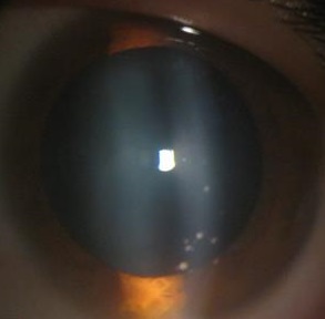
References
Urrego-DÃaz JA, FrÃas-Ordoñez JS, Figueroa-EchandÃa G, Durán-Silva G. Acute corneal edema without epithelium compromise. A case report and literature review. Rev Fac Med. 2017;65(3):513-9. doi: 10.15446/revfacmed.v65n3.56637
Koizumi N, Suzuki T, Uno T, Chihara H, Shiraishi A, Hara Y, et al. Cytomegalovirus as an etiologic factor in corneal endotheliitis. Ophthalmology. 2008;115(2):292-7 e3. doi: 10.1016/j.ophtha.2007.04.053 pmid: 17669498
Eslani M, Baradaran-Rafii A, Movahedan A, Djalilian AR. The ocular surface chemical burns. J Ophthalmol. 2014;2014:196827. doi: 10.1155/2014/196827 pmid: 25105018
Fish R, Davidson RS. Management of ocular thermal and chemical injuries, including amniotic membrane therapy. Curr Opin Ophthalmol. 2010;21(4):317-21. doi: 10.1097/ICU.0b013e32833a8da2 pmid: 20467317
Madhavan HN, Goldsmith CS, Rao SK, Fogla R, Malathi J, Priya K. Isolation of a vesicular virus belonging to the family rhabdoviridae from the aqueous humor of a patient with bilateral corneal endotheliitis. Cornea. 2002;21(3):333-5. pmid: 11917189
Wilhelmus KR, Sugar J, Hyndiuk RA, Stulting RD. Corneal thickness changes during herpes simplex virus disciform keratitis. Cornea. 2004;23(2):154-7. pmid: 15075884
Ishikawa S, Kato N, Takeuchi M. Quantitative evaluation of corneal epithelial edema after cataract surgery using corneal densitometry: a prospective study. BMC Ophthalmol. 2018;18(1):334. doi: 10.1186/s12886-018-0998-5 pmid: 30572855
Elflein HM, Hofherr T, Berisha-Ramadani F, Weyer V, Lampe C, Beck M, et al. Measuring corneal clouding in patients suffering from mucopolysaccharidosis with the Pentacam densitometry programme. Br J Ophthalmol. 2013;97(7):829-33. doi: 10.1136/bjophthalmol-2012-302913 pmid: 23685998
Greenstein SA, Fry KL, Bhatt J, Hersh PS. Natural history of corneal haze after collagen crosslinking for keratoconus and corneal ectasia: Scheimpflug and biomicroscopic analysis. J Cataract Refract Surg. 2010;36(12):2105-14. doi: 10.1016/j.jcrs.2010.06.067 pmid: 21111314
Takacs AI, Mihaltz K, Nagy ZZ. Corneal density with the Pentacam after photorefractive keratectomy. J Refract Surg. 2011;27(4):269-77. doi: 10.3928/1081597X-20100618-02 pmid: 20672772
Al-Amry MA, Al-Ghadeer HA. Corneal Epitheliopathy After Trauma by Fake Snow Powder in a 7-year-old Child. Middle East Afr J Ophthalmol. 2016;23(3):274-6. doi: 10.4103/0974-9233.186157 pmid: 27555717
Levenson JE. Corneal edema: cause and treatment. Surv Ophthalmol. 1975;20(3):190-204. doi: 10.1016/0039-6257(75)90002-8 pmid: 1105846
Laganowski HC, Sherrard ES, Muir MG, Buckley RJ. Distinguishing features of the iridocorneal endothelial syndrome and posterior polymorphous dystrophy: value of endothelial specular microscopy. Br J Ophthalmol. 1991;75(4):212-6. doi: 10.1136/bjo.75.4.212 pmid: 2021586
Hamill CE, Schmedt T, Jurkunas U. Fuchs endothelial cornea dystrophy: a review of the genetics behind disease development. Semin Ophthalmol. 2013;28(5-6):281-6. doi: 10.3109/08820538.2013.825283 pmid: 24138036
Liesegang TJ. Classification of herpes simplex virus keratitis and anterior uveitis. Cornea. 1999;18(2):127-43. pmid: 10090358
Holland EJ, Schwartz GS. Classification of herpes simplex virus keratitis. Cornea. 1999;18(2):144-54. doi: 10.1097/00003226-199903000-00002 pmid: 10090359
Inoue Y. Review of clinical and basic approaches to corneal endotheliitis. Cornea. 2014;33 Suppl 11:S3-8. doi: 10.1097/ICO.0000000000000228 pmid: 25170583
Abulafia A, Segev F, Platner E, Ben Simon GJ. Party foam-induced eye injuries and the power of media intervention. Cornea. 2013;32(6):826-9. doi: 10.1097/ICO.0b013e31826cf315 pmid: 23132449
Kobayashi R, Hashida N, Soma T, Koh S, Miki A, Usui S, et al. Clinical Findings of Anterior Segment Spectral Domain Optical Coherence Tomography Images in Cytomegalovirus Corneal Endotheliitis. Cornea. 2017;36(4):411-4. doi: 10.1097/ICO.0000000000001103 pmid: 27941387
Cennamo G, Forte R, Aufiero B, La Rana A. Computerized Scheimpflug densitometry as a measure of corneal optical density after excimer laser refractive surgery in myopic eyes. J Cataract Refract Surg. 2011;37(8):1502-6. doi: 10.1016/j.jcrs.2011.03.037 pmid: 21782093
Ambrosio R, Jr., Valbon BF, Faria-Correia F, Ramos I, Luz A. Scheimpflug imaging for laser refractive surgery. Curr Opin Ophthalmol. 2013;24(4):310-20. doi: 10.1097/ICU.0b013e3283622a94 pmid: 23680761
Savini G, Barboni P, Carbonelli M, Hoffer KJ. Accuracy of corneal power measurements by a new Scheimpflug camera combined with Placido-disk corneal topography for intraocular lens power calculation in unoperated eyes. J Cataract Refract Surg. 2012;38(5):787-92. doi: 10.1016/j.jcrs.2011.11.037 pmid: 22386277
Peyman A, Pourazizi M. Scheimpflug-based Optical Densitometry for Assessment of Corneal Opacity: An Objective Method to Monitor Interstitial Keratitis. J Ophthalmic Vis Res. 2018;13(2):207-9. doi: 10.4103/jovr.jovr_96_17 pmid: 29719653
- Abstract Viewed: 1165 times
- Free Full Text PDF Downloaded: 765 times
