Comparison of Corneal Epithelial Thickness Map Measured by Spectral Domain Optical Coherence Tomography in Healthy, Subclinical and Early Keratoconus Subjects
Medical hypothesis discovery and innovation in ophthalmology,
Vol. 8 No. 2 (2019),
15 June 2019
,
Page 85-91
Abstract
The aim of this study was to compare epithelial thickness map obtained by Spectral Domain Optical Coherence Tomography (SD-OCT) of eyes with myopic astigmatism but without keratoconus, subclinical and early keratoconus. Sixty-three eyes divided into three groups; myopic astigmatism without keratoconus, subclinical and early keratoconus. Corneal epithelial thickness map was obtained by SD-OCT for all patients and compared between the 3 groups. Mean epithelial thickness in the area of minimum corneal epithelial thickness, in the one eighth part of the inferior (I) and in the one eighth part of the temporal (T) were 56.64±2.82 µm, 59.00±3.24 µm and 60.40±4.93 µm respectively in subclinical group. Three parameters on epithelial maps obtained by SD-OCT was significantly different in the 2 groups: I and T corneal epithelial thickness map was thicker in subclinical keratoconus (P<0.02 and P<0.02 respectively). Epithelial map uniformity indices were different between the groups, as Superior-I, Superonasal-Inferotemporal were lower (P<0.00 and P< 0.01 respectively) but T-nasal was higher in the subclinical group (P<0.02). The area with minimum epithelial thickness had a significantly lower amount in early keratoconus group compared to the other two groups (P<0.00). In conclusion, corneal epithelial thickness map provided early detection of keratoconus in the subclinical stage with compensatory epithelial thickening of inferior and temporal one eighth compared to total corneal thickness and changes in epithelial map uniformity indices cause early detection of subclinical keratoconus from normal cornea.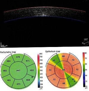
References
Rabinowitz YS. Keratoconus. Surv Ophthalmol. 1998;42(4):297-319. doi: 10.1016/s0039-6257(97)001 19-7 pmid: 9493273
Binder PS, Lindstrom RL, Stulting RD, Donnenfeld E, Wu H, McDonnell P, et al. Keratoconus and corneal ectasia after LASIK. J Cataract Refract Surg. 2005;31(11):2035-8. doi: 10.1016/j.jcrs.2005.12.002 pmid: 16412891
Kanellopoulos AJ. Collagen cross-linking in early keratoconus with riboflavin in a femtosecond laser-created pocket: initial clinical results. J Refract Surg. 2009;25(11):1034-7. doi: 10.3928/1081597X-200909 01-02 pmid: 19731884
Li Y, Chamberlain W, Tan O, Brass R, Weiss JL, Huang D. Subclinical keratoconus detection by pattern analysis of corneal and epithelial thickness maps with optical coherence tomography. J Cataract Refract Surg. 2016;42(2):284-95. doi: 10.1016/j.jcrs.2015.09.0 21 pmid: 27026454
Reinstein DZ, Archer TJ, Gobbe M. Corneal epithelial thickness profile in the diagnosis of keratoconus. J Refract Surg. 2009;25(7):604-10. pmid: 19662917
Avitabile T, Franco L, Ortisi E, Castiglione F, Pulvirenti M, Torrisi B, et al. Keratoconus staging: a computer-assisted ultrabiomicroscopic method compared with videokeratographic analysis. Cornea. 2004;23(7):655-60. pmid: 15448489
Li X, Rabinowitz YS, Rasheed K, Yang H. Longitudinal study of the normal eyes in unilateral keratoconus patients. Ophthalmology. 2004;111(3):440-6. doi: 10.1016/j.ophtha.2003.06.020 pmid: 15019316
Catalan S, Cadarso L, Esteves F, Salgado-Borges J, Lopez M, Cadarso C. Assessment of Corneal Epithelial Thickness in Asymmetric Keratoconic Eyes and Normal Eyes Using Fourier Domain Optical Coherence Tomography. J Ophthalmol. 2016;2016:5697343. doi: 10.1155/2016/5697343 pmid: 27379181
Ambrosio R, Jr., Dawson DG, Salomao M, Guerra FP, Caiado AL, Belin MW. Corneal ectasia after LASIK despite low preoperative risk: tomographic and biomechanical findings in the unoperated, stable, fellow eye. J Refract Surg. 2010;26(11):906-11. doi: 10.3928/1081597X-20100428-02 pmid: 20481412
Klein SR, Epstein RJ, Randleman JB, Stulting RD. Corneal ectasia after laser in situ keratomileusis in patients without apparent preoperative risk factors. Cornea. 2006;25(4):388-403. doi: 10.1097/01.ico.00 00222479.68242.77 pmid: 16670 474
Seiler T, Quurke AW. Iatrogenic keratectasia after LASIK in a case of forme fruste keratoconus. J Cataract Refract Surg. 1998;24(7):1007-9. doi: 10.1016/s0886-3350(98)80057-6 pmid: 9682124
Li Y, Tan O, Brass R, Weiss JL, Huang D. Corneal epithelial thickness mapping by Fourier-domain optical coherence tomography in normal and keratoconic eyes. Ophthalmology. 2012;119(12):2425-33. doi: 10.1016/j.ophtha.2012 .06.023 pmid: 22917888
Reinstein DZ, Silverman RH, Sutton HF, Coleman DJ. Very high-frequency ultrasound corneal analysis identifies anatomic correlates of optical complications of lamellar refractive surgery: anatomic diagnosis in lamellar surgery. Ophthalmology. 1999;106(3):474-82. doi: 10.1016/S0161-6420(99)90105-7 pmid: 10080202
Reinstein DZ, Archer T. Combined Artemis very high-frequency digital ultrasound-assisted transepithelial phototherapeutic keratectomy and wavefront-guided treatment following multiple corneal refractive procedures. J Cataract Refract Surg. 2006;32(11):1870-6. doi: 10.1016/j.jcrs.2006.07.016 pmid: 17081871
Reinstein DZ, Gobbe M, Archer TJ, Silverman RH, Coleman DJ. Epithelial, stromal, and total corneal thickness in keratoconus: three-dimensional display with artemis very-high frequency digital ultrasound. J Refract Surg. 2010;26(4):259-71. doi: 10.3928/1081597X-20100218-01 pmid: 20415322
Moller-Pedersen T, Vogel M, Li HF, Petroll WM, Cavanagh HD, Jester JV. Quantification of stromal thinning, epithelial thickness, and corneal haze after photorefractive keratectomy using in vivo confocal microscopy. Ophthalmology. 1997;104(3):360-8. doi: 10.1016/s0161-6420(97)30307-8 pmid: 9082257
Reinstein DZ, Archer TJ, Gobbe M, Silverman RH, Coleman DJ. Epithelial thickness in the normal cornea: three-dimensional display with Artemis very high-frequency digital ultrasound. J Refract Surg. 2008;24(6):571-81. doi: 10.3928/1081597X-20080601-05 pmid: 18581782
Huang D, Swanson EA, Lin CP, Schuman JS, Stinson WG, Chang W, et al. Optical coherence tomography. Science. 1991;254(5035):1178-81. pmid: 1957169
Krumeich JH, Daniel J, Knulle A. Live-epikeratophakia for keratoconus. J Cataract Refract Surg. 1998;24(4):456-63. doi: 10.1016/s0886-3350(98)802 84-8 pmid: 9584238
Elhennawi FM, Alzankalony YA, Abdellatif MK, Ibrahim AMT. Role of Anterior Segment Optical Coherence Tomography in The Diagnosis of Subclinical Keratoconus in Comparison with The Pentacam. Egypt J Hosp Med. 2018;72(1):3712-5.
Silverman RH, Urs R, Roychoudhury A, Archer TJ, Gobbe M, Reinstein DZ. Epithelial remodeling as basis for machine-based identification of keratoconus. Invest Ophthalmol Vis Sci. 2014;55(3):1580-7. doi: 10.1167/iovs.13-12578 pmid: 24557351
Sandali O, El Sanharawi M, Temstet C, Hamiche T, Galan A, Ghouali W, et al. Fourier-domain optical coherence tomography imaging in keratoconus: a corneal structural classification. Ophthalmology. 2013;120(12):2403-12. doi: 10.1016/j.ophtha.2013.0 5.027 pmid: 23932599
Sella R, Zangwill LM, Weinreb RN, Afshari NA. Repeatability and Reproducibility of Corneal Epithelial Thickness Mapping With Spectral-Domain Optical Coherence Tomography in Normal and Diseased Cornea Eyes. Am J Ophthalmol. 2019;197:88-97. doi: 10.1016/j.ajo.2018.09.008 pmid: 30240724
Tang M, Shekhar R, Miranda D, Huang D. Characteristics of keratoconus and pellucid marginal degeneration in mean curvature maps. Am J Ophthalmol. 2005 Dec;140(6):993-1001. pmid: 16376641
Temstet C, Sandali O, Bouheraoua N, Hamiche T, Galan A, El Sanharawi M, et al. Corneal epithelial thickness mapping using Fourier-domain optical coherence tomography for detection of form fruste keratoconus. J Cataract Refract Surg. 2015;41(4):812-20. doi: 10.1016/j.jcrs.2014.06.043 pmid: 25840306
Pflugfelder SC, Liu Z, Feuer W, Verm A. Corneal thickness indices discriminate between keratoconus and contact lens-induced corneal thinning. Ophthalmology. 2002;109(12):2336-41. doi: 10.1016/s0161-6420(02)01276-9 pmid: 12466180
Haque S, Jones L, Simpson T. Thickness mapping of the cornea and epithelium using optical coherence tomography. Optom Vis Sci. 2008;85(10):E963-76. pmid: 18832971
Prospero Ponce CM, Rocha KM, Smith SD, Krueger RR. Central and peripheral corneal thickness measured with optical coherence tomography, Scheimpflug imaging, and ultrasound pachymetry in normal, keratoconus-suspect, and post-laser in situ keratomileusis eyes. J Cataract Refract Surg. 2009 Jun;35(6):1055-62. doi: 10.1016/j.jcrs.2009.01.022. pmid: 19465292
- Abstract Viewed: 1078 times
- Free Full Text PDF Downloaded: 861 times
