Multimodal Imaging of Acute Central Retinal Artery Occlusion
Medical hypothesis discovery and innovation in ophthalmology,
Vol. 8 No. 4 (2019),
29 December 2019
,
Page 283-290
Abstract
The aim of this study was to describe fluorescein angiography (FA), ocular coherence tomography (OCT) and ocular coherence tomography angiography (OCTA) in the diagnosis of acute central retinal artery occlusion (CRAO). This is an observational case series study performed at Sohag Ophthalmic Investigation Center. Fifteen patients presented by a sudden marked unilateral diminution of vision were included. Corrected Distance Visual acuity (CDVA), color fundus photos, FA, OCT and OCTA, imaging obtained in the first week of presentation and imaging of the other normal eye as a control were assessed. Central macular thickness (CMT), parafoveal inner retinal layers thickness and parafoveal outer retinal thickness in diseased and contralateral normal eyes were compared. Fifteen patients (mean age 52.67 years, 11-74 years old) including 66.7% male entered the study. CDVA ranged from no perception of light to 0.05 (20/400). Fundus examination showed a cherry red spot in 10 cases (66.7 %) and retinal whitening in 9 cases (60%), arteriolar narrowing in 7 (46.67%), optic disc edema in 4 (26.67%), optic disc pallor in 5 (33.3%) and cattle trucking in 5 (33.3%). Fluorescein angiography showed delayed arteriovenous transit time > 23 seconds in 8 cases (53.33 %) and normal FA in 4 cases (26.67 %). OCT revealed increased hyperreflective of the inner retinal layers in comparison to hyporeflective inner retinal layers in all cases (100%) and significant increase in CMT in 10 cases (66.67%). The mean ± standard deviation (SD) of CMT (CRAO) was 306.5 ± 27.9 (P < 0.001), the parafoveal inner retinal thickness (CRAO) 345 ± 51.8 µm (P < 0.001) and the parafoveal outer retinal thickness (CRAO) 120.9 ± 13.6 µm (P < 0.001). OCTA was performed and clear images obtained in 11 cases (73.33%). Disruption of superficial and deep capillary plexus was found in all cases. We concluded that the OCT is the most confirmative imaging method in the diagnosis of acute CRAO even in the absence of fundus signs. OCTA confirms the diagnosis, but it cannot be performed in some cases. Epub: October 1, 2019.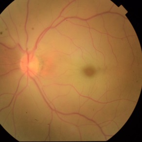
References
Hayreh SS. Ocular vascular occlusive disorders: natural history of visual outcome. Prog Retin Eye Res. 2014;41:1-25. doi: 10.1016/j.preteyeres.2014.04.001 pmid: 24769221
Varma DD, Cugati S, Lee AW, Chen CS. A review of central retinal artery occlusion: clinical presentation and management. Eye (Lond). 2013;27(6):688-97. doi: 10.1038/eye.2013.25 pmid: 23470793
Hayreh SS, Podhajsky PA, Zimmerman MB. Branch retinal artery occlusion: natural history of visual outcome. Ophthalmology. 2009;116(6):1188-94 e1-4. doi: 10.1016/j.ophtha.2009.01.015 pmid: 19376586
Hayreh SS. Acute retinal arterial occlusive disorders. Prog Retin Eye Res. 2011;30(5):359-94. doi: 10.1016/j.preteyeres.2011.05.001 pmid: 21620994
Mehta N, Marco RD, Goldhardt R, Modi Y. Central Retinal Artery Occlusion: Acute Management and Treatment. Curr Ophthalmol Rep. 2017;5(2):149-59. doi: 10.1007/s40135-017-0135-2 pmid: 29051845
Ghazi NG, Tilton EP, Patel B, Knape RM, Newman SA. Comparison of macular optical coherence tomography findings between postacute retinal artery occlusion and nonacute optic neuropathy. Retina. 2010;30(4):578-85. doi: 10.1097/IAE.0b013e3181bf5fd5 pmid: 19996826
Yu S, Wang F, Pang CE, Yannuzzi LA, Freund KB. Multimodal imaging findings in retinal deep capillary ischemia. Retina. 2014;34(4):636-46. doi: 10.1097/IAE.0000000000000048 pmid: 24240565
Gong H, Song Q, Wang L. Manifestations of central retinal artery occlusion revealed by fundus fluorescein angiography are associated with the degree of visual loss. Exp Ther Med. 2016;11(6):2420-4. doi: 10.3892/etm.2016.3175 pmid: 27313672
Ahn SJ, Woo SJ, Park KH, Jung C, Hong JH, Han MK. Retinal and choroidal changes and visual outcome in central retinal artery occlusion: an optical coherence tomography study. Am J Ophthalmol. 2015;159(4):667-76. doi: 10.1016/j.ajo.2015.01.001 pmid: 25579642
Matsunaga D, Yi J, Puliafito CA, Kashani AH. OCT angiography in healthy human subjects. Ophthalmic Surg Lasers Imaging Retina. 2014;45(6):510-5. doi: 10.3928/23258160-20141118-04 pmid: 25423629
Bonini Filho MA, Adhi M, de Carlo TE, Ferrara D, Baumal CR, Witkin AJ, et al. Optical Coherence Tomography Angiography in Retinal Artery Occlusion. Retina. 2015;35(11):2339-46. doi: 10.1097/IAE.0000000000000850 pmid: 26457398
Graefe A. Ueber Embolie der Arteria centralis retinae als Ursache plötzlicher Erblindung. Albrecht von Graefes Arch Ophthalmol. 1859;5(1):136-57. doi: 10.1007/bf02720764
Hayreh SS, Zimmerman MB. Central retinal artery occlusion: visual outcome. Am J Ophthalmol. 2005;140(3):376-91. doi: 10.1016/j.ajo.2005.03.038 pmid: 16138997
Furashova O, Matthe E. Retinal Changes in Different Grades of Retinal Artery Occlusion: An Optical Coherence Tomography Study. Invest Ophthalmol Vis Sci. 2017;58(12):5209-16. doi: 10.1167/iovs.17-22411 pmid: 29049721
Hayreh SS, Zimmerman MB. Fundus changes in central retinal artery occlusion. Retina. 2007;27(3):276-89. doi: 10.1097/01.iae.0000238095.97104.9b pmid: 17460582
Hayreh SS, Zimmerman MB, Kimura A, Sanon A. Central retinal artery occlusion. Retinal survival time. Exp Eye Res. 2004;78(3):723-36. doi: 10.1016/s0014-4835(03)00214-8 pmid: 15106952
Schmidt DP, Schulte-Monting J, Schumacher M. Prognosis of central retinal artery occlusion: local intraarterial fibrinolysis versus conservative treatment. AJNR Am J Neuroradiol. 2002;23(8):1301-7. pmid: 12223369
Schmidt D, Schumacher M. Stage-dependent efficacy of intra-arterial fibrinolysis in central retinal artery occlusion (CRAO). Neuro-Ophthalmol. 2009;20(3):125-41. doi: 10.1076/noph.20.3.125.4155
Dotan G, Goldenberg D, Kesler A, Naftaliev E, Loewenstein A, Goldstein M. The use of spectral-domain optical coherence tomography for differentiating long-standing central retinal artery occlusion and nonarteritic anterior ischemic optic neuropathy. Ophthalmic Surg Lasers Imaging Retina. 2014;45(1):38-44. doi: 10.3928/23258160-20131220-05 pmid: 24392910
Cornut PL, Bieber J, Beccat S, Fortoul V, Poli M, Feldman A, et al. [Spectral domain OCT in eyes with retinal artery occlusion]. J Fr Ophtalmol. 2012;35(8):606-13. doi: 10.1016/j.jfo.2012.04.008 pmid: 22819341
Ozdemir H, Karacorlu S, Karacorlu M. Optical coherence tomography findings in central retinal artery occlusion. Retina. 2006;26(1):110-2. pmid: 16395151
Chen SN, Hwang JF, Chen YT. Macular thickness measurements in central retinal artery occlusion by optical coherence tomography. Retina. 2011;31(4):730-7. doi: 10.1097/IAE.0b013e3181f2a15c pmid: 21242861
Yanoff M. Retinal Ischemia. Ocular Pathology-A Text And Atlas. Philadelphia: Lippincott Co; 1989.
Feucht N, Zapp D, Reznicek L, Lohmann CP, Maier M, Mayer CS. Multimodal imaging in acute retinal ischemia: spectral domain OCT, OCT-angiography and fundus autofluorescence. Int J Ophthalmol. 2018;11(9):1521-7. doi: 10.18240/ijo.2018.09.15 pmid: 30225228
Lee AY, Zhang Q, Baughman DM, Mudumbai R, Wang RK, Lee CS. Evaluation of bilateral central retinal artery occlusions with optical coherence tomography-based microangiography: a case report. J Med Case Rep. 2016;10(1):307. doi: 10.1186/s13256-016-1095-0 pmid: 27802835
Hwang CK, Kolomeyer AM, Brucker AJ. Optical Coherence Tomography Angiography of a Central Retinal Artery Occlusion Before and After Anterior Chamber Paracentesis. Ophthalmology. 2017;124(5):608. doi: 10.1016/j.ophtha.2016.10.030 pmid: 28433123
Philippakis E, Dupas B, Bonnin P, Hage R, Gaudric A, Tadayoni R. Optical Coherence Tomography Angiography Shows Deep Capillary Plexus Hypoperfusion in Incomplete Central Retinal Artery Occlusion. Retin Cases Brief Rep. 2015;9(4):333-8. doi: 10.1097/ICB.0000000000000211 pmid: 26355822
Shah P, Schwartz SG, Flynn HW, Jr. Multimodal Images of Acute Central Retinal Artery Occlusion. Case Rep Ophthalmol Med. 2017;2017:5151972. doi: 10.1155/2017/5151972 pmid: 29348953
Damento G, Chen MH, Leng T. Spectral-Domain Optical Coherence Tomography Angiography of Central Retinal Artery Occlusion. Ophthalmic Surg Lasers Imaging Retina. 2016;47(5):467-70. doi: 10.3928/23258160-20160419-10 pmid: 27183551
Bhanushali DR, Yadav NK, Dabir S, Chidambara L, Srinivasan P, Shetty R. Spectral Domain Optical Coherence Tomography Angiography Features in a Patient of Central Retinal Arterial Occlusion Before and After Paracentesis. Retina. 2016;36(5):e36-8. doi: 10.1097/IAE.0000000000000908 pmid: 26756807
Kwiterovich KA, Maguire MG, Murphy RP, Schachat AP, Bressler NM, Bressler SB, et al. Frequency of adverse systemic reactions after fluorescein angiography. Results of a prospective study. Ophthalmology. 1991;98(7):1139-42. doi: 10.1016/s0161-6420(91)32165-1 pmid: 1891225
- Abstract Viewed: 1157 times
- Full Text PDF. Epub:Oct. 1, 2019 Downloaded: 546 times
