Use of Fundus Autofluorescence Combined with Optical Coherence Tomography for Diagnose of Geographic Atrophy in Age-Related Macular Degeneration
Medical hypothesis discovery and innovation in ophthalmology,
Vol. 8 No. 4 (2019),
29 December 2019
,
Page 298-305
Abstract
The aim of this study was to demonstrate the sensitivity of Optical coherence tomography (OCT) in detection of geographic atrophy (GA) secondary to exudative age related macular degeneration (AMD). In this retrospective case series study 77 patients (53% female, with mean ± standard deviation [SD] of 82.6±9.3 years) with 97 eyes (45 OS [left eyes]/52 OD [right eyes]) were included. This was a retrospective review of the charts of patients who presented with exudative AMD at the Pitié Salpetrière Hospital, Paris, France, between December 2016 and August 2017 that received intravitreal injections of anti-vascular endothelial growth factor (anti-VEGF) therapies. At baseline, following biomicroscopy examination, multimodal imaging was performed including, fluorescein angiography (FA), fundus auto-fluorescence (FAF), spectral domain optical coherence tomography (SD-OCT) and indocyanine green angiography (ICGA). During the follow-up, SD-OCT with/without FAF and FA were performed for each patient at 6, 12 and 18 months. For investigation of the prevalence of GA in eyes undergoing intravitreal injections with anti-VEGF therapy, FAF and SD-OCT images were qualitatively reviewed by four independent observers (two graders per group). Kappa coefficient of Cohen was calculated to determine agreement between the graders. The kappa coefficient of Cohen, for inter-rater agreement in the evaluation of FAF images was 0.468, indicating a moderate agreement between the first and second raters. Thus, the sensitivity and specificity of FAF for the diagnosis of GA were 70% and 57%, respectively. If atrophy was assessed with SD-OCT image analysis, the kappa coefficient for inter-rater agreement was 0.846, implying an acceptable agreement between both readers. The sensitivity and specificity of SD-OCT were 93% and 58% respectively. In conclusion, SD-OCT image analysis was more sensitive than FAF for identifying GA in patients treated for exudative AMD. Epub: October 1, 2019.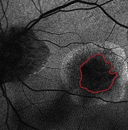
References
Bressler NM. Age-related macular degeneration is the leading cause of blindness. JAMA. 2004;291(15):1900-1. doi: 10.1001/jama.291.15.1900 pmid: 15108691
Ferris FL, Fine SL, Hyman L. Age-related macular degeneration and blindness due to neovascular maculopathy. Arch Ophthalmol. 1984;102(11):1640-2. doi: 10.1001/archopht.1984.01040031330019 pmid: 6208888
Hubschman JP, Reddy S, Schwartz SD. Age-related macular degeneration: current treatments. Clin Ophthalmol. 2009;3:155-66. doi: 10.2147/opth.s2094 pmid: 19668560
ANAES report. Dégénérescence maculaire liée à l'âge: prise en charge diagnostique et thérapeutique. ANAES 2015.
Investigators IS, Chakravarthy U, Harding SP, Rogers CA, Downes SM, Lotery AJ, et al. Ranibizumab versus bevacizumab to treat neovascular age-related macular degeneration: one-year findings from the IVAN randomized trial. Ophthalmology. 2012;119(7):1399-411. doi: 10.1016/j.ophtha.2012.04.015 pmid: 22578446
Group CR, Martin DF, Maguire MG, Ying GS, Grunwald JE, Fine SL, et al. Ranibizumab and bevacizumab for neovascular age-related macular degeneration. N Engl J Med. 2011;364(20):1897-908. doi: 10.1056/NEJMoa1102673 pmid: 21526923
Comparison of Age-related Macular Degeneration Treatments Trials Research G, Martin DF, Maguire MG, Fine SL, Ying GS, Jaffe GJ, et al. Ranibizumab and bevacizumab for treatment of neovascular age-related macular degeneration: two-year results. Ophthalmology. 2012;119(7):1388-98. doi: 10.1016/j.ophtha.2012.03.053 pmid: 22555112
Chakravarthy U, Harding SP, Rogers CA, Downes S, Lotery AJ, Dakin HA, et al. A randomised controlled trial to assess the clinical effectiveness and cost-effectiveness of alternative treatments to Inhibit VEGF in Age-related choroidal Neovascularisation (IVAN). Health Technol Assess. 2015;19(78):1-298. doi: 10.3310/hta19780 pmid: 26445075
Frank G, Holz M, Erich C, Strauss M, Steffen Schmitz-Valckenberg M. Geographic Atrophy. Clin Features Potent Therapeut Appr. 2014.
Mata NL, Lichter JB, Vogel R, Han Y, Bui TV, Singerman LJ. Investigation of oral fenretinide for treatment of geographic atrophy in age-related macular degeneration. Retina. 2013;33(3):498-507. doi: 10.1097/IAE.0b013e318265801d pmid: 23023528
Holz FG, Bindewald-Wittich A, Fleckenstein M, Dreyhaupt J, Scholl HP, Schmitz-Valckenberg S, et al. Progression of geographic atrophy and impact of fundus autofluorescence patterns in age-related macular degeneration. Am J Ophthalmol. 2007;143(3):463-72. doi: 10.1016/j.ajo.2006.11.041 pmid: 17239336
Klein R, Meuer SM, Knudtson MD, Klein BE. The epidemiology of progression of pure geographic atrophy: the Beaver Dam Eye Study. Am J Ophthalmol. 2008;146(5):692-9. doi: 10.1016/j.ajo.2008.05.050 pmid: 18672224
Klein R, Davis MD, Magli YL, Segal P, Klein BE, Hubbard L. The Wisconsin age-related maculopathy grading system. Ophthalmology. 1991;98(7):1128-34. doi: 10.1016/s0161-6420(91)32186-9 pmid: 1843453
Rofagha S, Bhisitkul RB, Boyer DS, Sadda SR, Zhang K, Group S-US. Seven-year outcomes in ranibizumab-treated patients in ANCHOR, MARINA, and HORIZON: a multicenter cohort study (SEVEN-UP). Ophthalmology. 2013;120(11):2292-9. doi: 10.1016/j.ophtha.2013.03.046 pmid: 23642856
Garrity ST, Sarraf D, Freund KB, Sadda SR. Multimodal Imaging of Nonneovascular Age-Related Macular Degeneration. Invest Ophthalmol Vis Sci. 2018;59(4):AMD48-AMD64. doi: 10.1167/iovs.18-24158 pmid: 30025107
Singer MA, Awh CC, Sadda S, Freeman WR, Antoszyk AN, Wong P, et al. HORIZON: an open-label extension trial of ranibizumab for choroidal neovascularization secondary to age-related macular degeneration. Ophthalmology. 2012;119(6):1175-83. doi: 10.1016/j.ophtha.2011.12.016 pmid: 22306121
Bird AC, Bressler NM, Bressler SB, Chisholm IH, Coscas G, Davis MD, et al. An international classification and grading system for age-related maculopathy and age-related macular degeneration. The International ARM Epidemiological Study Group. Surv Ophthalmol. 1995;39(5):367-74. doi: 10.1016/s0039-6257(05)80092-x pmid: 7604360
Grossniklaus HE, Martinez JA, Brown VB, Lambert HM, Sternberg P, Jr., Capone A, Jr., et al. Immunohistochemical and histochemical properties of surgically excised subretinal neovascular membranes in age-related macular degeneration. Am J Ophthalmol. 1992;114(4):464-72. doi: 10.1016/s0002-9394(14)71859-8 pmid: 1415458
Das A, Puklin JE, Frank RN, Zhang NL. Ultrastructural immunocytochemistry of subretinal neovascular membranes in age-related macular degeneration. Ophthalmology. 1992;99(9):1368-76. doi: 10.1016/s0161-6420(92)31792-0 pmid: 1407971
Ishikawa K, Kannan R, Hinton DR. Molecular mechanisms of subretinal fibrosis in age-related macular degeneration. Exp Eye Res. 2016;142:19-25. doi: 10.1016/j.exer.2015.03.009 pmid: 25773985
Jager RD, Mieler WF, Miller JW. Age-related macular degeneration. N Engl J Med. 2008;358(24):2606-17. doi: 10.1056/NEJMra0801537 pmid: 18550876
Ferris FL, 3rd, Wilkinson CP, Bird A, Chakravarthy U, Chew E, Csaky K, et al. Clinical classification of age-related macular degeneration. Ophthalmology. 2013;120(4):844-51. doi: 10.1016/j.ophtha.2012.10.036 pmid: 23332590
Jang KH, Do YJ, Son D, Son E, Choi JS, Kim E. AIF-independent parthanatos in the pathogenesis of dry age-related macular degeneration. Cell Death Dis. 2017;8(1):e2526. doi: 10.1038/cddis.2016.437 pmid: 28055012
Du J, Yanagida A, Knight K, Engel A, Vo A, Jankowski C, et al. Reductive carboxylation is a noteworthy metabolic pathway in the retinal shade epithelium. Proc Natl Acad Sci. 2016;113(51):14710-5.
Bonilha VL. Age and disease-related structural changes in the retinal pigment epithelium. Clin Ophthalmol. 2008;2(2):413-24. doi: 10.2147/opth.s2151 pmid: 19668732
Querques L, Parravano M, Borrelli E, Chiaravalloti A, Tedeschi M, Sacconi R, et al. Anatomical and practical changes in neovascular AMD disappearing: correlation of fibrocellular and fibrovascular phenotypes. B J Ophthalmol. 2018;8(5):31-6.
Bhakhri R. Spectral domain optical coherence tomography and auto-fluorescence findings in adult-onset vitelliform dystrophy. Clin Exp Optom. 2015;98(3):292-3. doi: 10.1111/cxo.12234 pmid: 25557869
Singh AD, Belfort RN, Sayanagi K, Kaiser PK. Fourier domain optical coherence tomographic and auto-fluorescence findings in indeterminate choroidal melanocytic lesions. Br J Ophthalmol. 2010;94(4):474-8. doi: 10.1136/bjo.2009.162636 pmid: 19822920
Holz FG, Steinberg JS, Gobel A, Fleckenstein M, Schmitz-Valckenberg S. Fundus autofluorescence imaging in dry AMD: 2014 Jules Gonin lecture of the Retina Research Foundation. Graefes Arch Clin Exp Ophthalmol. 2015;253(1):7-16. doi: 10.1007/s00417-014-2858-1 pmid: 25408425
Chen Q, de Sisternes L, Leng T, Zheng L, Kutzscher L, Rubin DL. Semi-automatic geographic atrophy segmentation for SD-OCT images. Biomed Opt Express. 2013;4(12):2729-50. doi: 10.1364/BOE.4.002729 pmid: 24409376
- Abstract Viewed: 1188 times
- Full Text PDF. Epub:Oct. 1, 2019 Downloaded: 570 times
