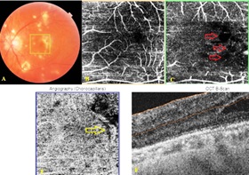
Optical Coherence Tomography Angiography in Eyes with Non-infectious Posterior Uveitis; Some Practical Aspects
Medical hypothesis discovery and innovation in ophthalmology,
Vol. 8 No. 4 (2019),
29 December 2019
,
Page 312-322
Abstract
Optical coherence tomography angiography (OCTA) is an innovative imaging technology enabling clinicians to learn more about the pathophysiology of disease processes as it facilitates visualization of the retinal and choroidal circulation without injection of a dye. Also it provides ample qualitative and quantitative data on the vascular supply. OCTA has become an important tool nowadays in the diagnosis and follow-up of patients with age-related macular degeneration, inherited chorioretinal diseases, diabetic retinopathy, retinal vascular occlusive diseases and optic nerve disorders. However, its place is relatively less known in non-infectious posterior uveitis (NIPU). OCTA may help mainly in assessing macular and peripheric retinal perfusion status, detection of retinal and/or disc neovascularization, diagnose of inflammatory choroidal neovascularization and visualizing the uveitic white-dot lesions. This mini-review describes the use of OCTA in patients with NIPU and summarizes some practical points in several uveitic entities. Epub: October 1, 2019.
References
Durrani OM, Tehrani NN, Marr JE, Moradi P, Stavrou P, Murray PI. Degree, duration, and causes of visual loss in uveitis. Br J Ophthalmol. 2004;88(9):1159-62. doi: 10.1136/bjo.2003.037226 pmid: 15317708
Mikhail M, Sallam A. Novel Intraocular Therapy in Non-infectious Uveitis of the Posterior Segment of the Eye. Med Hypothesis Discov Innov Ophthalmol. 2013;2(4):113-20. pmid: 24822232
Karti O, Saatci AO. Intravitreal Dexamethasone Implant in the Treatment of Non-Infectious Uveitic Macular Edema. Med Hypothesis Discov Innov Ophthalmol. 2018;7(4):169-75. pmid: 30505868
Smit RL, Baarsma GS. Epidemiology of uveitis. Curr Opin Ophthalmol. 1995;6(3):57-61. pmid: 10150871
London NJ, Rathinam SR, Cunningham ET, Jr. The epidemiology of uveitis in developing countries. Int Ophthalmol Clin. 2010;50(2):1-17. doi: 10.1097/IIO.0b013e3181d2cc6b pmid: 20375859
Miserocchi E, Fogliato G, Modorati G, Bandello F. Review on the worldwide epidemiology of uveitis. Eur J Ophthalmol. 2013;23(5):705-17. doi: 10.5301/ejo.5000278 pmid: 23661536
Moschos MM. Subclinical Macular Edema Detected by Spectral-domain Optical Coherence Tomography (SD-OCT) in HLA-B27 Positive Anterior Uveitis. Med Hypothesis Discov Innov Ophthalmol. 2014;3(4):123-6. pmid: 25756062
Suttorp-Schulten MS, Rothova A. The possible impact of uveitis in blindness: a literature survey. Br J Ophthalmol. 1996;80(9):844-8. doi: 10.1136/bjo.80.9.844 pmid: 8962842
de Smet MD, Taylor SR, Bodaghi B, Miserocchi E, Murray PI, Pleyer U, et al. Understanding uveitis: the impact of research on visual outcomes. Prog Retin Eye Res. 2011;30(6):452-70. doi: 10.1016/j.preteyeres.2011.06.005 pmid: 21807112
Spaide RF, Klancnik JM, Jr., Cooney MJ. Retinal vascular layers in macular telangiectasia type 2 imaged by optical coherence tomographic angiography. JAMA Ophthalmol. 2015;133(1):66-73. doi: 10.1001/jamaophthalmol.2014.3950 pmid: 25317692
Coscas G, Lupidi M, Coscas F. Image Analysis of Optical Coherence Tomography Angiography. Dev Ophthalmol. 2016;56:30-6. doi: 10.1159/000442774 pmid: 27023365
de Carlo TE, Romano A, Waheed NK, Duker JS. A review of optical coherence tomography angiography (OCTA). Int J Retina Vitreous. 2015;1:5. doi: 10.1186/s40942-015-0005-8 pmid: 27847598
Ipek SC, Ayhan Z, Kadayifcilar S, Saatci AO. Swept-source Optical Coherence Tomography Angiography in a Patient with Bietti Crystalline Dystrophy Followed for Ten Years. Turk J Ophthalmol. 2019;49(2):106-8. doi: 10.4274/tjo.galenos.2018.90768 pmid: 31055897
Borrelli E, Sarraf D, Freund KB, Sadda SR. OCT angiography and evaluation of the choroid and choroidal vascular disorders. Prog Retin Eye Res. 2018;67:30-55. doi: 10.1016/j.preteyeres.2018.07.002 pmid: 30059755
Li XX, Wu W, Zhou H, Deng JJ, Zhao MY, Qian TW, et al. A quantitative comparison of five optical coherence tomography angiography systems in clinical performance. Int J Ophthalmol. 2018;11(11):1784-95. doi: 10.18240/ijo.2018.11.09 pmid: 30450309
Rodriguez FJ, Staurenghi G, Gale R, Vision Academy Steering C. The role of OCT-A in retinal disease management. Graefes Arch Clin Exp Ophthalmol. 2018;256(11):2019-26. doi: 10.1007/s00417-018-4109-3 pmid: 30175386
Munk MR, Giannakaki-Zimmermann H, Berger L, Huf W, Ebneter A, Wolf S, et al. OCT-angiography: A qualitative and quantitative comparison of 4 OCT-A devices. PLoS One. 2017;12(5):e0177059. doi: 10.1371/journal.pone.0177059 pmid: 28489918
Zhang A, Zhang Q, Chen CL, Wang RK. Methods and algorithms for optical coherence tomography-based angiography: a review and comparison. J Biomed Opt. 2015;20(10):100901. doi: 10.1117/1.JBO.20.10.100901 pmid: 26473588
Kashani AH, Chen CL, Gahm JK, Zheng F, Richter GM, Rosenfeld PJ, et al. Optical coherence tomography angiography: A comprehensive review of current methods and clinical applications. Prog Retin Eye Res. 2017;60:66-100. doi: 10.1016/j.preteyeres.2017.07.002 pmid: 28760677
Zhang Q, Zheng F, Motulsky EH, Gregori G, Chu Z, Chen CL, et al. A Novel Strategy for Quantifying Choriocapillaris Flow Voids Using Swept-Source OCT Angiography. Invest Ophthalmol Vis Sci. 2018;59(1):203-11. doi: 10.1167/iovs.17-22953 pmid: 29340648
Spaide RF, Fujimoto JG, Waheed NK. Image Artifacts in Optical Coherence Tomography Angiography. Retina. 2015;35(11):2163-80. doi: 10.1097/IAE.0000000000000765 pmid: 26428607
Miller AR, Roisman L, Zhang Q, Zheng F, Rafael de Oliveira Dias J, Yehoshua Z, et al. Comparison Between Spectral-Domain and Swept-Source Optical Coherence Tomography Angiographic Imaging of Choroidal Neovascularization. Invest Ophthalmol Vis Sci. 2017;58(3):1499-505. doi: 10.1167/iovs.16-20969 pmid: 28273316
Novais EA, Adhi M, Moult EM, Louzada RN, Cole ED, Husvogt L, et al. Choroidal Neovascularization Analyzed on Ultrahigh-Speed Swept-Source Optical Coherence Tomography Angiography Compared to Spectral-Domain Optical Coherence Tomography Angiography. Am J Ophthalmol. 2016;164:80-8. doi: 10.1016/j.ajo.2016.01.011 pmid: 26851725
Invernizzi A, Cozzi M, Staurenghi G. Optical coherence tomography and optical coherence tomography angiography in uveitis: A review. Clin Exp Ophthalmol. 2019;47(3):357-71. doi: 10.1111/ceo.13470 pmid: 30719788
Lipson BK, Yannuzzi LA. Complications of intravenous fluorescein injections. Int Ophthalmol Clin. 1989;29(3):200-5. doi: 10.1097/00004397-198902930-00011 pmid: 2526795
Cerquaglia A, Iaccheri B, Fiore T, Fruttini D, Belli FB, Khairallah M, et al. New Insights On Ocular Sarcoidosis: An Optical Coherence Tomography Angiography Study. Ocul Immunol Inflamm. 2018:1-10. doi: 10.1080/09273948.2018.1497665 pmid: 30081683
Pichi F, Srvivastava SK, Chexal S, Lembo A, Lima LH, Neri P, et al. EN FACE OPTICAL COHERENCE TOMOGRAPHY AND OPTICAL COHERENCE TOMOGRAPHY ANGIOGRAPHY OF MULTIPLE EVANESCENT WHITE DOT SYNDROME: New Insights Into Pathogenesis. Retina. 2016;36 Suppl 1:S178-S88. doi: 10.1097/IAE.0000000000001255 pmid: 28005676
Baxter SL, Pistilli M, Pujari SS, Liesegang TL, Suhler EB, Thorne JE, et al. Risk of choroidal neovascularization among the uveitides. Am J Ophthalmol. 2013;156(3):468-77 e2. doi: 10.1016/j.ajo.2013.04.040 pmid: 23795984
Roy R, Saurabh K, Bansal A, Kumar A, Majumdar AK, Paul SS. Inflammatory choroidal neovascularization in Indian eyes: Etiology, clinical features, and outcomes to anti-vascular endothelial growth factor. Indian J Ophthalmol. 2017;65(4):295-300. doi: 10.4103/ijo.IJO_262_16 pmid: 28513493
D'Ambrosio E, Tortorella P, Iannetti L. Management of uveitis-related choroidal neovascularization: from the pathogenesis to the therapy. J Ophthalmol. 2014;2014:450428. doi: 10.1155/2014/450428 pmid: 24868454
Gass JD. Pathogenesis of tears of the retinal pigment epithelium. Br J Ophthalmol. 1984;68(8):513-9. doi: 10.1136/bjo.68.8.513 pmid: 6204685
Grossniklaus HE, Gass JD. Clinicopathologic correlations of surgically excised type 1 and type 2 submacular choroidal neovascular membranes. Am J Ophthalmol. 1998;126(1):59-69. doi: 10.1016/s0002-9394(98)00145-7 pmid: 9683150
Agarwal A, Invernizzi A, Singh RB, Foulsham W, Aggarwal K, Handa S, et al. An update on inflammatory choroidal neovascularization: epidemiology, multimodal imaging, and management. J Ophthalmic Inflamm Infect. 2018;8(1):13. doi: 10.1186/s12348-018-0155-6 pmid: 30209691
Astroz P, Miere A, Mrejen S, Sekfali R, Souied EH, Jung C, et al. Optical Coherence Tomography Angiography to Distinguish Choroidal Neovascularization from Macular Inflammatory Lesions in Multifocal Choroiditis. Retina. 2018;38(2):299-309. doi: 10.1097/IAE.0000000000001617 pmid: 28368976
Nozaki M, Hamada S, Kimura M, Yoshida M, Ogura Y. Value of OCT Angiography in the Diagnosis of Choroidal Neovascularization Complicating Multiple Evanescence White Dot Syndrome. Ophthalmic Surg Lasers Imaging Retina. 2016;47(6):580-4. doi: 10.3928/23258160-20160601-11 pmid: 27327289
Levison AL, Baynes KM, Lowder CY, Kaiser PK, Srivastava SK. Choroidal neovascularisation on optical coherence tomography angiography in punctate inner choroidopathy and multifocal choroiditis. Br J Ophthalmol. 2017;101(5):616-22. doi: 10.1136/bjophthalmol-2016-308806 pmid: 27539089
Baumal CR, de Carlo TE, Waheed NK, Salz DA, Witkin AJ, Duker JS. Sequential Optical Coherence Tomographic Angiography for Diagnosis and Treatment of Choroidal Neovascularization in Multifocal Choroiditis. JAMA Ophthalmol. 2015;133(9):1087-90. doi: 10.1001/jamaophthalmol.2015.1946 pmid: 26111344
Nakao S, Kaizu Y, Oshima Y, Sakamoto T, Ishibashi T, Sonoda KH. Optical Coherence Tomography Angiography for Detecting Choroidal Neovascularization Secondary to Punctate Inner Choroidopathy. Ophthalmic Surg Lasers Imaging Retina. 2016;47(12):1157-61. doi: 10.3928/23258160-20161130-13 pmid: 27977842
Fujiwara A, Morizane Y, Hosokawa M, Kimura S, Shiode Y, Hirano M, et al. Factors affecting foveal avascular zone in healthy eyes: An examination using swept-source optical coherence tomography angiography. PLoS One. 2017;12(11):e0188572. doi: 10.1371/journal.pone.0188572 pmid: 29176837
Waizel M, Todorova MG, Terrada C, LeHoang P, Massamba N, Bodaghi B. Superficial and deep retinal foveal avascular zone OCTA findings of non-infectious anterior and posterior uveitis. Graefes Arch Clin Exp Ophthalmol. 2018;256(10):1977-84. doi: 10.1007/s00417-018-4057-y pmid: 29980918
Cheng D, Shen M, Zhuang X, Lin D, Dai M, Chen S, et al. Inner Retinal Microvasculature Damage Correlates With Outer Retinal Disruption During Remission in Behcet's Posterior Uveitis by Optical Coherence Tomography Angiography. Invest Ophthalmol Vis Sci. 2018;59(3):1295-304. doi: 10.1167/iovs.17-23113 pmid: 29625452
Khairallah M, Abroug N, Khochtali S, Mahmoud A, Jelliti B, Coscas G, et al. Optical Coherence Tomography Angiography in Patients with Behcet Uveitis. Retina. 2017;37(9):1678-91. doi: 10.1097/IAE.0000000000001418 pmid: 28002270
Ahn SJ, Park SH, Lee BR. Multimodal Imaging Including Optical Coherence Tomography Angiography in Serpiginous Choroiditis. Ocul Immunol Inflamm. 2017;25(2):287-91. doi: 10.1080/09273948.2017.1288824 pmid: 28281863
Aggarwal K, Agarwal A, Mahajan S, Invernizzi A, Mandadi SKR, Singh R, et al. The Role of Optical Coherence Tomography Angiography in the Diagnosis and Management of Acute Vogt-Koyanagi-Harada Disease. Ocul Immunol Inflamm. 2018;26(1):142-53. doi: 10.1080/09273948.2016.1195001 pmid: 27440118
Heiferman MJ, Rahmani S, Jampol LM, Nesper PL, Skondra D, Kim LA, et al. Acute Posterior Multifocal Placoid Pigment Epitheliopathy on Optical Coherence Tomography Angiography. Retina. 2017;37(11):2084-94. doi: 10.1097/IAE.0000000000001487 pmid: 28151840
Burke TR, Chu CJ, Salvatore S, Bailey C, Dick AD, Lee RWJ, et al. Application of OCT-angiography to characterise the evolution of chorioretinal lesions in acute posterior multifocal placoid pigment epitheliopathy. Eye (Lond). 2017;31(10):1399-408. doi: 10.1038/eye.2017.180 pmid: 28983094
Klufas MA, Phasukkijwatana N, Iafe NA, Prasad PS, Agarwal A, Gupta V, et al. Optical Coherence Tomography Angiography Reveals Choriocapillaris Flow Reduction in Placoid Chorioretinitis. Ophthalmol Retina. 2017;1(1):77-91. doi: 10.1016/j.oret.2016.08.008 pmid: 31047399
Forte R, Saleh M, Aptel F, Chiquet C. Evaluation of Photoreceptors, Retinal Capillary Plexuses, and Choriocapillaris in Patients with Birdshot Chorioretinopathy. Retina. 2019. doi: 10.1097/IAE.0000000000002457 pmid: 30789462
de Carlo TE, Bonini Filho MA, Adhi M, Duker JS. Retinal and Choroidal Vasculature in Birdshot Chorioretinopathy Analyzed Using Spectral Domain Optical Coherence Tomography Angiography. Retina. 2015;35(11):2392-9. doi: 10.1097/IAE.0000000000000744 pmid: 26352557
Roberts PK, Nesper PL, Goldstein DA, Fawzi AA. Retinal Capillary Density in Patients with Birdshot Chorioretinopathy. Retina. 2018;38(2):387-94. doi: 10.1097/IAE.0000000000001543 pmid: 28196058
Hassan M, Agarwal A, Afridi R, daSilva MJ, Karaca I, Sadiq MA, et al. The Role of Optical Coherence Tomography Angiography in the Management of Uveitis. Int Ophthalmol Clin. 2016;56(4):1-24. doi: 10.1097/IIO.0000000000000130 pmid: 27575755
Dingerkus VLS, Munk MR, Brinkmann MP, Freiberg FJ, Heussen FMA, Kinzl S, et al. Optical coherence tomography angiography (OCTA) as a new diagnostic tool in uveitis. J Ophthalmic Inflamm Infect. 2019;9(1):10. doi: 10.1186/s12348-019-0176-9 pmid: 31139955
Park JJ, Soetikno BT, Fawzi AA. Characterization of the Middle Capillary Plexus Using Optical Coherence Tomography Angiography in Healthy and Diabetic Eyes. Retina. 2016;36(11):2039-50. doi: 10.1097/IAE.0000000000001077 pmid: 27205895
Nemiroff J, Kuehlewein L, Rahimy E, Tsui I, Doshi R, Gaudric A, et al. Assessing Deep Retinal Capillary Ischemia in Paracentral Acute Middle Maculopathy by Optical Coherence Tomography Angiography. Am J Ophthalmol. 2016;162:121-32 e1. doi: 10.1016/j.ajo.2015.10.026 pmid: 26562176
Leder HA, Campbell JP, Sepah YJ, Gan T, Dunn JP, Hatef E, et al. Ultra-wide-field retinal imaging in the management of non-infectious retinal vasculitis. J Ophthalmic Inflamm Infect. 2013;3(1):30. doi: 10.1186/1869-5760-3-30 pmid: 23514542
Doganay N, Balikoglu Yilmaz M, Orduyilmaz B, Aydin E, Saatci AO. Central Serous Chorioretinopathy: A Complication Associated with Behcet's Disease Treatment. Turk J Ophthalmol. 2019;49(1):40-3. doi: 10.4274/tjo.galenos.2018.83479 pmid: 30829024
Guyer DR, Schachat AP, Green WR. The Choroid: Structural Considerations. In: Ryan S, Schachat A, Wilkinson C, Hinton D, editors. Retina. Edinburgh, Scotland: Mosby; 2006. p. 33-42.
Mullins RF, Johnson MN, Faidley EA, Skeie JM, Huang J. Choriocapillaris vascular dropout related to density of drusen in human eyes with early age-related macular degeneration. Invest Ophthalmol Vis Sci. 2011;52(3):1606-12. doi: 10.1167/iovs.10-6476 pmid: 21398287
McLeod DS, Grebe R, Bhutto I, Merges C, Baba T, Lutty GA. Relationship between RPE and choriocapillaris in age-related macular degeneration. Invest Ophthalmol Vis Sci. 2009;50(10):4982-91. doi: 10.1167/iovs.09-3639 pmid: 19357355
Mullins RF, Schoo DP, Sohn EH, Flamme-Wiese MJ, Workamelahu G, Johnston RM, et al. The membrane attack complex in aging human choriocapillaris: relationship to macular degeneration and choroidal thinning. Am J Pathol. 2014;184(11):3142-53. doi: 10.1016/j.ajpath.2014.07.017 pmid: 25204844
Spaide RF, Fujimoto JG, Waheed NK, Sadda SR, Staurenghi G. Optical coherence tomography angiography. Prog Retin Eye Res. 2018;64:1-55. doi: 10.1016/j.preteyeres.2017.11.003 pmid: 29229445
Cole ED, Moult EM, Dang S, Choi W, Ploner SB, Lee B, et al. The Definition, Rationale, and Effects of Thresholding in OCT Angiography. Ophthalmol Retina. 2017;1(5):435-47. doi: 10.1016/j.oret.2017.01.019 pmid: 29034359
- Abstract Viewed: 1425 times
- Full Text PDF. Epub:Oct. 1, 2019 Downloaded: 656 times


