Multimodal Imaging Characteristics of a Large Retinal Capillary Macroaneurysm in an Eye With Severe Diabetic Macular Edema: A Case Presentation and Literature Review
Medical hypothesis discovery and innovation in ophthalmology,
Vol. 9 No. 1 (2020),
19 November 2019
,
Page 33-37
Abstract
Though microaneurysms are the hallmark of diabetic retinopathy (DR), large aneurismal changes termed as ''macroaneurysms'' (MAs) may also occur in the course of chronic diabetic macular edema. MAs are usually accompanied by intraretinal hard exudates, fluid accumulation and retinal hemorrhages. Detection of MAs is clinically important as it implies that macular edema is usually chronic and therefore can be resistant to intravitreal anti-vascular endothelial growth factor injections. Multimodal imaging consisting of fluorescein angiography (FA), indocyanine green angiography (ICGA), optical coherence tomography (OCT) or OCT-angiography (OCTA) can be performed to detect and understand the nature of MA and thereby select proper treatment modality. Herein, we report multimodal imaging features of a 64-year-old woman with insulin-dependent diabetes mellitus presented with treatment naive severe macular edema and a macroaneurysm at the right temporal macula. In conclusion, FA, ICGA and OCT seem to be far superior to OCTA to detect these lesions due to probable slow flow inside the MA.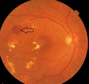
References
Barot M, Gokulgandhi MR, Patel S, Mitra AK. Microvascular complications and diabetic retinopathy: recent advances and future implications. Future Med Chem. 2013;5(3):301-14. doi: 10.4155/fmc.12.206 pmid: 23464520
Bourhis A, Girmens JF, Boni S, Pecha F, Favard C, Sahel JA, et al. Imaging of macroaneurysms occurring during retinal vein occlusion and diabetic retinopathy by indocyanine green angiography and high resolution optical coherence tomography. Graefes Arch Clin Exp Ophthalmol. 2010;248(2):161-6. doi: 10.1007/s00417-009-1175-6 pmid: 19701812
Stitt AW, Gardiner TA, Archer DB. Histological and ultrastructural investigation of retinal microaneurysm development in diabetic patients. Br J Ophthalmol. 1995;79(4):362-7. doi: 10.1136/bjo.79.4.362 pmid: 7742285
Castro Farias D, Matsui Serrano R, Bianchi Gancharov J, de Dios Cuadras U, Sahel J, Graue Wiechers F, et al. Indocyanine green angiography for identifying telangiectatic capillaries in diabetic macular oedema. Br J Ophthalmol. 2019. doi: 10.1136/bjophthalmol-2019-314355 pmid: 31358497
Paques M, Philippakis E, Bonnet C, Falah S, Ayello-Scheer S, Zwillinger S, et al. Indocyanine-green-guided targeted laser photocoagulation of capillary macroaneurysms in macular oedema: a pilot study. Br J Ophthalmol. 2017;101(2):170-4. doi: 10.1136/bjophthalmol-2015-308142 pmid: 27267449
Hamada M, Ohkoshi K, Inagaki K, Ebihara N, Murakami A. Visualization of microaneurysms using optical coherence tomography angiography: comparison of OCTA en face, OCT B-scan, OCT en face, FA, and IA images. Jpn J Ophthalmol. 2018;62(2):168-75. doi: 10.1007/s10384-018-0570-0 pmid: 29383540
Demir G, Artunay O, Sucu ME, Demircan A, Yasa D, Alagoz C, et al. Treatment of intravitreal bevacizumab combined with focal laser photocoagulation in the case of macular telangiectasia type 2 with retinal arterial macroaneurysm. Lasers Med Sci. 2019;34(1):235-8. doi: 10.1007/s10103-018-2548-z pmid: 29804166
Mansour AM, Foster RE, Gallego-Pinazo R, Moschos MM, Sisk RA, Chhablani J, et al. Intravitreal Anti-Vascular Endothelial Growth Factor Injections for Exudative Retinal Arterial Macroaneurysms. Retina. 2019;39(6):1133-41. doi: 10.1097/IAE.0000000000002131 pmid: 29505440
Cahuzac A, Scemama C, Mauget-Faysse M, Sahel JA, Wolff B. Retinal arterial macroaneurysms: clinical, angiographic, and tomographic description and therapeutic management of a series of 14 cases. Eur J Ophthalmol. 2016;26(1):36-43. doi: 10.5301/ejo.5000641 pmid: 26165327
Chanana B, Azad RV. Intravitreal bevacizumab for macular edema secondary to retinal macroaneurysm. Eye (Lond). 2009;23(2):493-4. doi: 10.1038/eye.2008.98 pmid: 18388958
Moser LA, Simpson DE, Young DD. Retinal macroaneurysms: the natural history in four patients. Optom Vis Sci. 1989;66(12):877-83. doi: 10.1097/00006324-198912000-00013 pmid: 2626256
Spaide RF, Barquet LA. Retinal Capillary Macroaneurysms. Retina. 2019;39(10):1889-95. doi: 10.1097/IAE.0000000000002406 pmid: 30489449
de Carlo TE, Romano A, Waheed NK, Duker JS. A review of optical coherence tomography angiography (OCTA). Int J Retina Vitreous. 2015;1:5. doi: 10.1186/s40942-015-0005-8 pmid: 27847598
Venkatesh R, Yadav NK, Bavaharan B, Prabhu V, Sinha S. Multimodal imaging in perifoveal exudative vascular anomalous complex with co-existent diabetic retinopathy. Clin Exp Optom. 2019;102(5):528-32. doi: 10.1111/cxo.12868 pmid: 30620998
Sacconi R, Freund KB, Yannuzzi LA, Dolz-Marco R, Souied E, Capuano V, et al. The Expanded Spectrum of Perifoveal Exudative Vascular Anomalous Complex. Am J Ophthalmol. 2017;184:137-46. doi: 10.1016/j.ajo.2017.10.009 pmid: 29079450.
- Abstract Viewed: 662 times
- Free Full Text PDF Downloaded: 526 times
